AdvertisementAdvertisement
Learn medical imaging, case by case
Teaching cases across six body systems, plus anatomy references and knowledge notes — structured, searchable and free for education.
StructuredHistory, findings, differential
AnnotatedEvery figure labelled
Six systemsNeuro to breast imaging
FreeFor teaching
Featured Cases
FEATURED CASE · Musculoskeletal
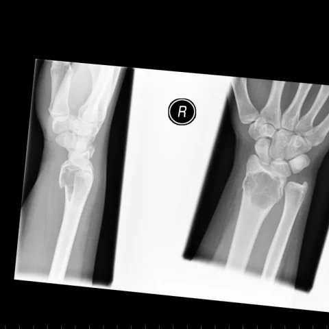
Giant cell tumour
Radiographs of the distal radius show an eccentric, expansile, lytic lesion extending to the subarticular surface of the distal radius, with thinned cortex and…
FEATURED CASE · Head and Neck
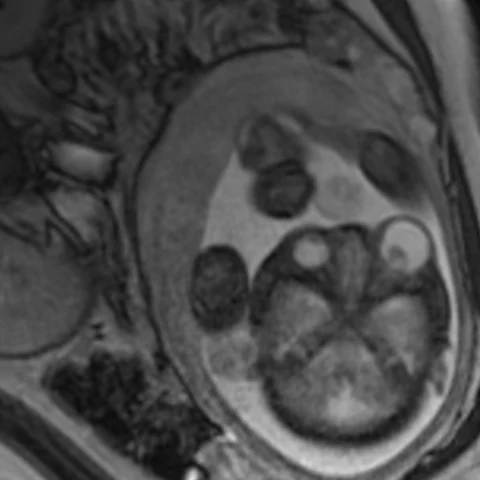
Congenital orbital teratoma
Fetal MRI shows a large, well-defined left orbital mass with both cystic and solid components, producing marked proptosis and displacement of the globe, and…
Latest from each system
View all →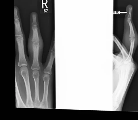 17 images
Intra-articular osteoid osteoma of the proximal phalanx
 3 images
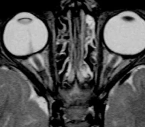
Persistent hyperplastic primary vitreous
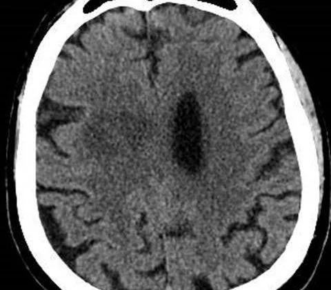 21 images
Pyogenic brain abscess with intraventricular rupture
 4 images
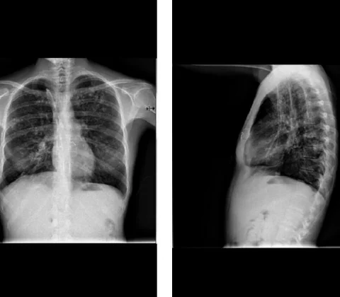
Metastatic pulmonary calcification secondary to haemodialysis
 3 images
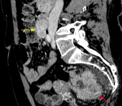
Synchronous duodenal GIST and colorectal adenocarcinoma
 8 images
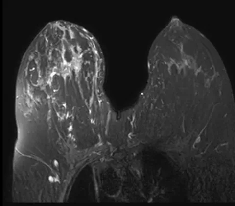
Multifocal high-grade DCIS with invasion
Browse by Modality
Browse by Topic
AdvertisementAdvertisement