Synchronous duodenal GIST and colorectal adenocarcinoma
Written and reviewed by RadLearn editorial team · Updated 20 Oct 2026
Figures (3)
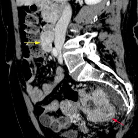
Patient history
A 53-year-old woman was admitted reporting asthma, pallor, recurrent rectal bleeding and constipation. Blood tests showed a low haemoglobin concentration (7.6 mg/dl). Digital rectal examination and clinical examination showed no pathological findings. She underwent CT to exclude a bleeding source.
Imaging findings — key points
- CT demonstrates a well-defined, lobulated, exophytic mass arising from the duodenal wall, with heterogeneous enhancement and central low-attenuation necrosis.
- A second, separate lesion is present in the sigmoid colon or rectum, showing irregular circumferential wall thickening with narrowing of the lumen and surrounding fat stranding.
- Enlarged regional lymph nodes are present in the mesocolon, and the staging CT shows no distant hepatic or pulmonary metastasis.
- The duodenal mass does not obstruct the bile duct or pancreatic duct and shows no calcification.
- Coronal reformats confirm the two tumours are anatomically separate, one in the duodenum and one in the colorectum.
Discussion
Gastrointestinal stromal tumours are mesenchymal neoplasms of the gastrointestinal tract that most often arise in the stomach and small bowel, and they typically present as well-circumscribed, exophytic, enhancing masses with central necrosis. Synchronous occurrence with a colorectal adenocarcinoma is uncommon but recognised, particularly in older patients, and each tumour must be staged and treated on its own merits. CT is the mainstay for detection, characterisation and staging, and it distinguishes a GIST from carcinoid, sarcoma and other submucosal lesions. Surgical resection is the treatment of choice for localised GIST, with tyrosine kinase inhibitors for advanced disease, while the colorectal cancer requires oncological and surgical management.
Differential diagnosis
| Alternative diagnosis | Key features that distinguish it from this case |
|---|---|
| Carcinoid tumour | A carcinoid is usually small, intensely enhancing and associated with mesenteric desmoplastic reaction and calcification, rather than a large necrotic exophytic duodenal mass. |
| Gastrointestinal sarcoma | A high-grade sarcoma is large and heterogeneous with necrosis and haemorrhage, but the presence of two distinct primary tumours makes a synchronous second primary more likely than a single sarcoma. |
| Leiomyosarcoma | Leiomyosarcoma arises from smooth muscle, often in the small bowel, and shows marked necrosis and heterogeneous enhancement, but it does not explain the separate colonic adenocarcinoma. |
| Gastrointestinal haemangioma | A haemangioma shows strong, persistent enhancement with phleboliths and no central necrosis, and it is not associated with a synchronous colonic tumour. |
The confirmed diagnosis itself is not included as an alternative — it is stated separately below.