Multifocal high-grade DCIS with invasion
Written and reviewed by RadLearn editorial team · Updated 20 Oct 2026
Figures (8)
Patient history
A 53-year-old postmenopausal woman presented for mammography due to occasional fullness in the right breast and a perceived size difference over several months. No palpable mass was clinically detected.
Imaging findings — key points
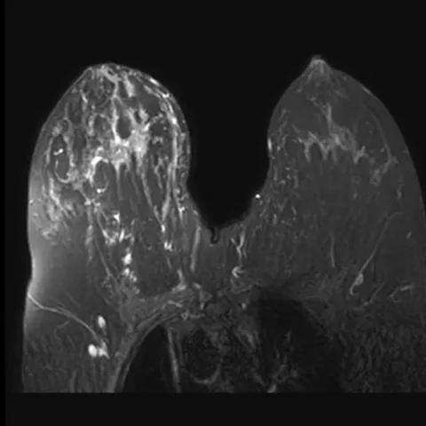
- Fat-suppressed post-contrast T1-weighted MRI shows multicentric areas of segmental, clumped non-mass enhancement in the right breast.
- Post-contrast maximum-intensity-projection images demonstrate several separate enhancing foci distributed across more than one quadrant.
- Ultrasound shows corresponding hypoechoic, ill-defined areas matching the enhancing regions.
- Craniocaudal and mediolateral oblique mammograms show a dense breast with developing asymmetry and possible pleomorphic microcalcification.
- The enhancement pattern is segmental and ductal rather than that of a solitary mass, without skin thickening or chest wall invasion.
Discussion
Multicentric high-grade DCIS with microinvasion is often mammographically subtle and is best delineated by contrast-enhanced MRI, which shows segmental clumped non-mass enhancement distributed across more than one quadrant. The extent of disease determines the surgical approach, and MRI is valuable for detecting multicentric foci before surgery. Ultrasound-guided or MRI-guided biopsy is required to confirm high-grade DCIS and to identify any invasive component.
Differential diagnosis
| Alternative diagnosis | Key features that distinguish it from this case |
|---|---|
| Invasive lobular carcinoma | Shows subtle ill-defined enhancement with architectural distortion and is often mammographically occult, unlike the ductal segmental calcification pattern. |
| Mastitis | Produces diffuse skin thickening, oedema and clustered ring enhancement with clinical inflammation rather than ductal non-mass enhancement. |
The confirmed diagnosis itself is not included as an alternative — it is stated separately below.