Pyogenic brain abscess with intraventricular rupture
Written and reviewed by RadLearn editorial team · Updated 20 Oct 2026
Figures (21)
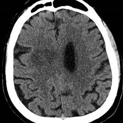
Patient history
A 49-year-old man with a history of alcohol abuse presented to the emergency department with fever, headache and cough over the previous four days. Confusion and slowed movements worsened his clinical condition. There was no relevant past medical history. Laboratory analysis showed leukocytosis and a raised C-reactive protein.
Imaging findings — key points
- Unenhanced CT shows an ill-defined hypodense area in the cerebral hemisphere with surrounding low-attenuation oedema and mass effect; contrast-enhanced CT demonstrates a thin, regular rim of enhancement around a hypodense necrotic centre.
- On MRI the cavity contents are markedly hyperintense on T2-weighted and FLAIR images, with high signal on DWI and low signal on the ADC map, indicating restricted diffusion of pus within the abscess core.
- The abscess capsule is hypointense on T2-weighted and susceptibility-weighted images and shows a dual-rim sign, with a hypointense outer collagenous rim and a hyperintense inner granulation-tissue rim.
- Extensive perilesional vasogenic oedema is present, extending through the white matter and producing sulcal effacement and midline shift.
- After intraventricular rupture there is dependent layering of debris in the occipital horns with FLAIR hyperintensity and restricted diffusion within the ventricular system, together with ventricular enlargement and ependymal enhancement after contrast administration.
Discussion
Pyogenic brain abscess develops from a focal cerebritis that evolves through early and late encephalitic stages into a encapsulated collection of pus, most often spread contiguously from paranasal sinus or dental infection, haematogenously, or after neurosurgery and trauma. Imaging findings mirror this evolution: an ill-defined area of oedema with patchy enhancement in early cerebritis and a thin, regular enhancing capsule with a restricted-diffusion core once encapsulation occurs. Recognising the capsule and its dual-rim sign separates an abscess from necrotic tumour, which is important because treatment differs. Intraventricular rupture is a feared complication that doubles mortality, and the sudden clinical deterioration with layering of pus in the dependent ventricles on FLAIR and DWI should prompt urgent neurosurgical and antimicrobial management.
Differential diagnosis
| Alternative diagnosis | Key features that distinguish it from this case |
|---|---|
| Metastasis | A metastasis usually arises at the grey-white matter junction with marked oedema, but its rim is thicker and more irregular and the central necrosis does not show the low ADC value of pus. |
| Glioblastoma | Glioblastoma shows an irregular, thick and nodular enhancing rim with central necrosis and increased perfusion, whereas a pyogenic abscess has a smooth thin capsule, restricted diffusion of the core and a dual-rim sign. |
| Tumour radionecrosis | Radionecrosis follows radiotherapy, shows a soap-bubble or Swiss-cheese enhancement pattern and has no restricted diffusion of the central contents. |
| Fungal, parasitic or mycobacterial abscess | These infections often show multiple lesions, a thicker or irregular capsule and specific features such as a scolex or tuberculoma-like nodule, and require microbiological correlation. |
The confirmed diagnosis itself is not included as an alternative — it is stated separately below.