Congenital orbital teratoma
Written and reviewed by RadLearn editorial team · Updated 20 Oct 2026
Figures (11)
Patient history
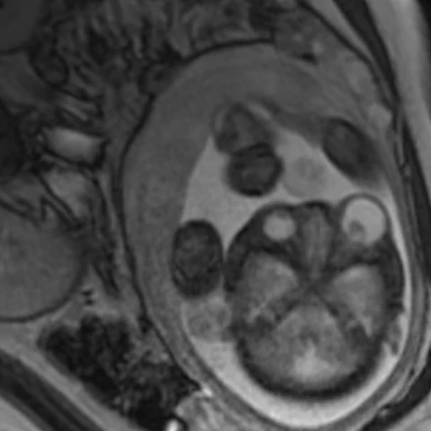
A left cystic orbital mass was detected incidentally on fetal ultrasound. Because of limited views on ultrasound, fetal MRI was performed to assess tumour extension. After delivery, examination showed a large erythematous cystic mass protruding from the left orbit, with no ocular structures. The infant underwent surgery at six months of age.
Imaging findings — key points
- Fetal MRI shows a large, well-defined left orbital mass with both cystic and solid components, producing marked proptosis and displacement of the globe, and extending towards the intracranial compartment without frank intracranial invasion.
- The mass is heterogeneous on T2-weighted images with multilocular hyperintense cysts, and it contains areas of fat signal and susceptibility artefact consistent with calcification.
- Postnatal CT confirms a heterogeneous orbital mass with fat-density areas, coarse calcification and bony remodelling of the orbital walls; no normal left globe is identified.
- The clinical photograph shows a large erythematous cystic mass protruding from the left orbit.
- Follow-up MRI after surgical excision shows removal of the mass with a residual orbital cavity and no recurrence.
Discussion
Congenital orbital teratoma is a rare germ-cell tumour that contains derivatives of all three germ layers and usually presents at birth with severe unilateral proptosis and a large cystic-solid orbital mass. Fetal MRI is valuable for defining the extent of the tumour and planning delivery, while postnatal CT and MRI characterise fat, calcification and cystic components that distinguish a teratoma from other congenital orbital lesions. The main differentials are congenital cystic eye, microphthalmos with cyst and retinoblastoma, none of which contains fat and calcification within a disorganised mass. Treatment is surgical excision, usually in the first months of life, and the prognosis depends on the degree of orbital and ocular destruction.
Differential diagnosis
| Alternative diagnosis | Key features that distinguish it from this case |
|---|---|
| Congenital cystic eye | Congenital cystic eye is a pure cyst with no solid component, fat or calcification and represents complete failure of globe formation, whereas a teratoma contains tissues from multiple germ layers. |
| Retinoblastoma | Retinoblastoma arises within a formed globe, shows intraocular calcification and does not produce a large extraocular mass with fat and cystic components. |
| Microphthalmos with cyst | Microphthalmos with cyst consists of a small globe continuous with a retrobulbar cyst, without solid enhancing tissue or calcification. |
| Orbital encephalocoele | An encephalocoele herniates through a bony defect and contains brain tissue with cerebrospinal fluid, and it is continuous with the intracranial compartment rather than a multilineage tumour. |
The confirmed diagnosis itself is not included as an alternative — it is stated separately below.