Giant cell tumour
Written and reviewed by RadLearn editorial team · Updated 20 Oct 2026
Figures (5)
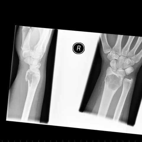
Patient history
A 14-year-old girl presented with right wrist pain after a fall. The injury was closed and neurovascular status was intact. Initial radiographs were obtained and the patient was treated in a cast. There was no pre-injury history of bone pain. The patient underwent curettage and cementation with more than five years of follow-up.
Imaging findings — key points
- Radiographs of the distal radius show an eccentric, expansile, lytic lesion extending to the subarticular surface of the distal radius, with thinned cortex and no sclerotic margin.
- CT confirms cortical thinning and focal breakthrough without internal calcification or ossification, and it shows the lesion abutting the articular surface.
- MRI demonstrates low to intermediate signal on T1-weighted images and heterogeneous high signal on T2-weighted images, with areas of fluid-fluid level consistent with secondary aneurysmal bone cyst change.
- The solid components enhance after gadolinium, and there is surrounding marrow and soft-tissue oedema.
- Follow-up radiographs after curettage and cementation show cement filling the cavity, and later images show callus formation after fixation of a pathological fracture.
Discussion
Giant cell tumour is a locally aggressive benign bone tumour that typically arises in the epiphysis of a long bone after skeletal maturity, most often around the knee and the distal radius. It is composed of mononuclear stromal cells and osteoclast-like giant cells and produces an eccentric, lytic, expansile lesion that reaches the subarticular surface without a sclerotic rim. MRI is important for defining the extent, detecting cortical breakthrough and identifying secondary aneurysmal bone cyst change with fluid-fluid levels, and the main differentials are aneurysmal bone cyst, enchondroma and simple bone cyst. Treatment is curettage with cementation or bone grafting, and recurrence occurs in a significant minority, so regular follow-up is required.
Differential diagnosis
| Alternative diagnosis | Key features that distinguish it from this case |
|---|---|
| Aneurysmal bone cyst | An aneurysmal bone cyst occurs in a younger patient, is metaphyseal and contains multiple fluid-fluid levels without solid enhancing tissue, whereas a giant cell tumour is epiphyseal and solid. |
| Enchondroma | An enchondroma is centrally located, contains chondroid ring-and-arc calcification and does not extend to the subarticular surface or enhance avidly. |
| Simple bone cyst | A simple bone cyst is central, fluid-filled and typically occurs in the proximal humerus or femur of a child, with no solid enhancing component. |
| Intraosseous ganglion | An intraosseous ganglion is a small, well-defined subarticular cyst with fluid signal and a sclerotic rim, without the solid enhancing tissue of a giant cell tumour. |
The confirmed diagnosis itself is not included as an alternative — it is stated separately below.