Persistent hyperplastic primary vitreous
Written and reviewed by RadLearn editorial team · Updated 20 Oct 2026
Figures (3)
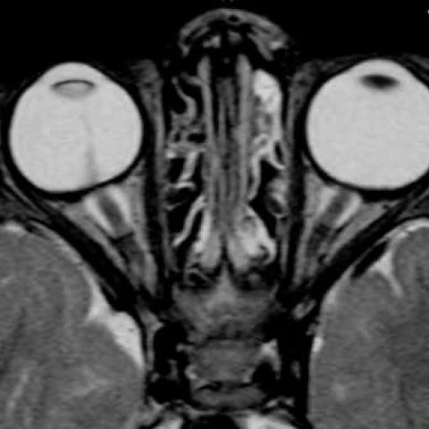
Patient history
Leukocoria with cataract formation, reduced vision in the right eye and delayed motor development.
Imaging findings — key points
- MRI of the orbits shows a unilateral microphthalmic globe with a retrolental soft-tissue mass and a thickened, hyperintense lens, consistent with cataract and persistent fetal lens remnants.
- A fibrous stalk extends from the posterior aspect of the lens to the optic disc along the expected course of the hyaloid canal, and it enhances after gadolinium administration.
- The retrolental mass is hyperintense on T2-weighted images and shows enhancement, and there is associated retinal detachment with a V-shaped configuration.
- No intraocular calcification is identified, and the retrobulbar optic nerve and extraocular muscles appear normal.
- The contralateral globe is normal in size and signal, supporting a unilateral developmental anomaly.
Discussion
Persistent hyperplastic primary vitreous, now called persistent fetal vasculature, is a congenital anomaly caused by failure of regression of the hyaloid vascular system, and it usually presents with leukocoria in a full-term infant. The eye is microphthalmic, and a retrolental mass with a stalk running to the optic disc, cataract and retinal detachment are the characteristic findings; contrast enhancement of the stalk helps to confirm the diagnosis. The main clinical concern is differentiation from retinoblastoma, which is important because the management and prognosis are entirely different, and the absence of calcification and the presence of a microphthalmic globe favour PHPV. Early surgery aims to clear the visual axis and prevent glaucoma and amblyopia.
Differential diagnosis
| Alternative diagnosis | Key features that distinguish it from this case |
|---|---|
| Retinoblastoma | Retinoblastoma typically arises in a normal-sized globe, contains calcification in most cases and forms an irregular intraocular mass without a persistent hyaloid stalk. |
| Coats disease | Coats disease causes exudative retinal detachment with hyperintense subretinal fluid and no retrolental mass or persistent hyaloid band, and it usually affects older boys. |
| Congenital cystic eye | Congenital cystic eye is a complete failure of globe formation with a cystic orbital mass and no identifiable eye, whereas in PHPV a microphthalmic globe is present. |
| Ocular toxocariasis | Toxocariasis produces a focal retinal granuloma with surrounding vitreous stranding and a history of contact with dogs or cats, without the congenital hyaloid stalk. |
The confirmed diagnosis itself is not included as an alternative — it is stated separately below.