Metastatic pulmonary calcification secondary to haemodialysis
Written and reviewed by RadLearn editorial team · Updated 20 Oct 2026
Figures (4)
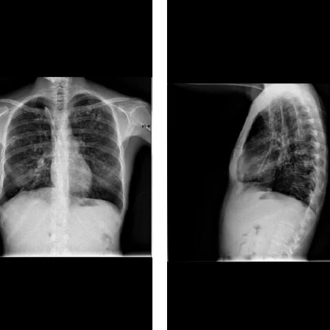
Patient history
A 45-year-old woman with polycystic kidney disease started a haemodialysis programme because of chronic renal failure. A chest radiograph showed ill-defined small nodules in the upper and mid lung zones.
Imaging findings — key points
- The chest radiograph shows ill-defined small nodules in the upper and middle lung zones, without cardiomegaly or pleural effusion.
- High-resolution CT demonstrates numerous centrilobular nodules with fluffy, poorly defined margins in the upper and mid zones, some of which are confluent.
- The nodules are densely calcified on the mediastinal window, with attenuation approaching that of bone, and there is no cavitation or surrounding ground-glass halo.
- The distribution spares the lung bases and the subpleural regions, and there is no lymphadenopathy or pleural thickening.
- No evidence of a primary neoplasm or granulomatous disease is seen on the available images.
Discussion
Metastatic pulmonary calcification is deposition of calcium in normal lung parenchyma in patients with a raised calcium-phosphate product, most often chronic renal failure treated with haemodialysis, and it may also occur in hyperparathyroidism, multiple myeloma or massive osteolysis. The deposits favour the upper and middle zones and appear as centrilobular fluffy nodules that calcify densely, and they are usually asymptomatic even when extensive. The main differential is alveolar microlithiasis, which is diffuse and basal, and other causes of calcified nodules such as prior granulomatous disease. Recognition matters because the condition is reversible in its early stages if the calcium-phosphate product is corrected, and it can progress to restrictive lung disease.
Differential diagnosis
| Alternative diagnosis | Key features that distinguish it from this case |
|---|---|
| Primary and secondary hyperparathyroidism | Hyperparathyroidism alone does not usually produce dense pulmonary nodular calcification, and the biochemical profile in this patient on haemodialysis points to metastatic calcification rather than a parathyroid adenoma. |
| Tuberous sclerosis | Tuberous sclerosis produces thin-walled lung cysts and multifocal micronodular pneumocyte hyperplasia with ground-glass nodules, not densely calcified upper-zone nodules. |
| Sarcoidosis | Sarcoidosis shows perilymphatic nodules along the bronchovascular bundles and fissures with bilateral hilar lymphadenopathy, rather than centrilobular calcified nodules. |
| Alveolar microlithiasis | Alveolar microlithiasis produces a diffuse sand-like micronodular pattern with basal predominance and a black pleura sign, unlike the upper-zone distribution here. |
The confirmed diagnosis itself is not included as an alternative — it is stated separately below.