Intra-articular osteoid osteoma of the proximal phalanx
Written and reviewed by RadLearn editorial team · Updated 20 Oct 2026
Figures (17)
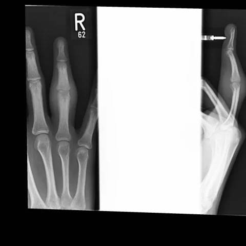
Patient history
A 30-year-old man had 2.5 years of painful swelling involving the proximal interphalangeal joint (PIPJ) of the right ring finger, with restricted movement. There was no previous injury and no exposure to tuberculosis (TB), but there was household contact with TB. Some relief was obtained with non-steroidal anti-inflammatory drugs (NSAIDs).
Imaging findings — key points
- The initial finger radiograph shows soft-tissue swelling around the proximal interphalangeal joint with mild joint space narrowing and periarticular sclerosis, but no obvious nidus.
- MRI demonstrates extensive bone marrow oedema in the proximal phalanx and middle phalanx, a joint effusion and synovial thickening, obscuring the underlying lesion.
- CT after joint fusion reveals a small, well-defined radiolucent nidus with central calcification in the subarticular bone of the proximal phalanx, surrounded by sclerosis.
- Power Doppler ultrasound shows hyperaemia around the affected joint, and MRI after fusion shows resolution of the marrow oedema.
- The final post-fusion radiograph shows a fused proximal interphalangeal joint with no residual lesion.
Discussion
Osteoid osteoma is a benign bone-forming tumour characterised by a small nidus of osteoid surrounded by reactive sclerosis, and when it lies within a joint it loses the typical sclerotic reaction and instead produces synovitis, effusion and joint contracture. Intra-articular lesions are frequently misdiagnosed as inflammatory or degenerative arthritis for months or years, and CT is the most reliable method for detecting the nidus. MRI is sensitive to the surrounding oedema but may obscure the nidus, and the clinical clue is pain that is worse at night and relieved by non-steroidal anti-inflammatory drugs. Treatment is radiofrequency ablation or surgical excision, and joint fusion is reserved for severe secondary arthropathy.
Differential diagnosis
| Alternative diagnosis | Key features that distinguish it from this case |
|---|---|
| Septic arthritis | Septic arthritis produces a hot, swollen joint with fever and rapid cartilage destruction, and it shows a joint effusion with synovial enhancement but no calcified nidus on CT. |
| Monoarticular arthropathy | Inflammatory or degenerative monoarthritis shows uniform joint space narrowing, erosions and synovial proliferation without a discrete nidus on CT. |
| Brodie abscess | A Brodie abscess has a central lytic area with a thick sclerotic rim and adjacent soft-tissue inflammation, and the patient usually has a history of infection and positive cultures. |
| Stress fracture | A stress fracture shows a linear fracture line with periosteal reaction and marrow oedema, without a rounded nidus containing calcification. |
The confirmed diagnosis itself is not included as an alternative — it is stated separately below.