Torsion of a wandering spleen
Written and reviewed by RadLearn editorial team · Updated 20 Oct 2026
Figures (5)
Patient history
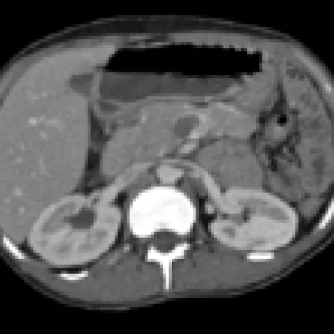
A 35-year-old woman was admitted with acute abdominal pain of 36 hours' duration. She had experienced similar episodes of pain over the past year. Her history was otherwise unremarkable. Physical examination found a large mass in the right abdominal quadrant with abdominal tenderness.
Imaging findings — key points
- Contrast-enhanced CT shows a large, well-defined mass in the right lower abdomen and pelvis that has the attenuation and enhancement pattern of splenic tissue.
- The spleen is absent from its normal position in the left upper quadrant, and the pancreatic tail is displaced medially.
- The splenic pedicle is elongated and shows a whirl sign of twisted vessels, with heterogeneous and reduced splenic enhancement indicating ischaemia.
- There is a small amount of perisplenic fluid and fat stranding, and no normal splenic tissue is seen in the left upper quadrant.
- Coronal reformats demonstrate the ectopic spleen occupying the right iliac fossa and crossing the midline, without invasion of adjacent organs.
Discussion
Wandering spleen results from laxity or absence of the suspensory ligaments that normally fix the spleen in the left upper quadrant, allowing it to migrate into the abdomen or pelvis, and it is a rare cause of acute abdominal pain when the elongated pedicle torts. Torsion produces venous congestion and arterial ischaemia, with an enlarged, heterogeneous, poorly enhancing spleen and a whirl sign of the twisted vessels, and it can progress to infarction. CT is the investigation of choice, showing the ectopic spleen, the empty splenic fossa and the twisted pedicle. Urgent detorsion with splenopexy is preferred when the spleen is viable, and splenectomy is required for infarction.
Differential diagnosis
| Alternative diagnosis | Key features that distinguish it from this case |
|---|---|
| Mesenteric cyst with small bowel volvulus | A mesenteric cyst is fluid-density with a thin wall and does not enhance like spleen, and the whirl sign involves bowel and mesentery rather than a splenic pedicle. |
| Ovarian torsion | Ovarian torsion produces an enlarged, oedematous adnexal mass with peripheral follicles and a twisted ovarian pedicle, and the spleen remains in its normal position. |
| Appendiceal mucocele | An appendiceal mucocele is a tubular cystic mass continuous with the caecum and does not show splenic enhancement or an absent spleen. |
| Renal mass | A renal mass arises from the kidney, shows renal enhancement and preserves a normal spleen in the left upper quadrant. |
The confirmed diagnosis itself is not included as an alternative — it is stated separately below.