Accessory spleen confirmed on histology
Written and reviewed by RadLearn editorial team · Updated 20 Oct 2026
Figures (5)
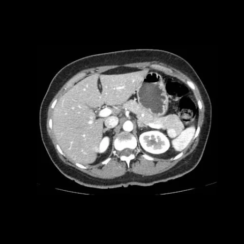
Patient history
A 48-year-old woman with persistent non-specific abdominal pain, arterial hypertension and depression. Surgical history included a left anterior resection. Laboratory tests showed no significant abnormalities.
Imaging findings — key points
- Axial CT in the hepatic arterial phase shows a well-defined, round mass in the left upper quadrant adjacent to the tail of the pancreas, with enhancement identical to that of the spleen.
- Curved planar reformats demonstrate a small vessel bridging the mass to the spleen, confirming its splenic origin.
- The mass follows the enhancement of the spleen in the portal venous phase, with no washout, capsule or central necrosis.
- The spleen is otherwise normal in size and appearance, and no other focal lesion is identified in the liver, pancreas or kidneys.
- There is no lymphadenopathy, ascites or vascular invasion.
Discussion
An accessory spleen is a common congenital variant resulting from failure of fusion of splenic tissue during embryogenesis, and it is usually an incidental finding in the splenic hilum or along the splenic vessels. The diagnosis is made when a well-defined mass enhances in parallel with the spleen on all phases and shows a bridging vessel, and no further investigation is needed. It matters mainly because it can be mistaken for a pancreatic or renal tumour, a lymph node or a metastasis, leading to unnecessary surgery. Recognition is also important in patients undergoing splenectomy, because an accessory spleen may hypertrophy and continue to cause symptoms in haematological disease.
Differential diagnosis
| Alternative diagnosis | Key features that distinguish it from this case |
|---|---|
| Papillary epithelial neoplasm | A solid pseudopapillary neoplasm of the pancreas is usually a large, heterogeneous cystic-solid mass in a young woman that enhances less than spleen and does not have a bridging vessel to the spleen. |
| Neuroendocrine tumour | A pancreatic neuroendocrine tumour is hypervascular in the arterial phase but shows washout and may be associated with hormonal symptoms, and it does not enhance in parallel with the spleen. |
| Vascular metastasis | A hypervascular metastasis shows arterial enhancement with washout, is often multiple and is associated with a known primary tumour. |
| Lymph node | An enlarged lymph node is homogeneous, has a fatty hilum and enhances less than spleen, without a bridging splenic vessel. |
The confirmed diagnosis itself is not included as an alternative — it is stated separately below.