Pancreatic lipoma
Written and reviewed by RadLearn editorial team · Updated 20 Oct 2026
Figures (5)
Patient history
A 90-year-old man was referred to the radiology department with upper abdominal pain persisting for one month.
Imaging findings — key points
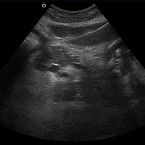
- Transabdominal ultrasound shows a well-defined, homogeneous, markedly hyperechoic mass within the pancreas, with no acoustic shadowing and no ductal dilatation.
- Axial contrast-enhanced CT demonstrates a round, well-circumscribed lesion in the pancreas with fat attenuation of approximately -90 to -110 HU, without enhancement or calcification.
- Axial T1- and T2-weighted MRI show the lesion to be hyperintense on both sequences, and it loses signal on fat-saturated images, confirming its fatty composition.
- The pancreatic duct and common bile duct are not dilated, and there is no peripancreatic stranding, lymphadenopathy or adjacent organ invasion.
- The remainder of the pancreas and the liver show normal attenuation and enhancement.
Discussion
Pancreatic lipoma is a rare benign tumour composed of mature adipocytes that is usually found incidentally and is asymptomatic, although large lesions may cause abdominal pain or obstructive symptoms. Imaging is diagnostic when a well-circumscribed mass with homogeneous fat attenuation on CT, hyperintensity on T1- and T2-weighted MRI and signal loss on fat saturation is seen, without enhancement or ductal obstruction. The main differentials are focal fatty infiltration, which is geographic and lacks a capsule, and pancreatic liposarcoma, which contains thick septa and solid enhancing tissue. No treatment is required for a typical lipoma, but follow-up or biopsy is warranted if the lesion is heterogeneous or grows.
Differential diagnosis
| Alternative diagnosis | Key features that distinguish it from this case |
|---|---|
| Focal fatty infiltration of the pancreas | Focal fatty infiltration is geographic, has no capsule or mass effect, follows the lobular architecture and shows normal enhancing pancreatic tissue within it. |
| Pancreatic liposarcoma | A liposarcoma is larger, heterogeneous with thick septa and nodular enhancing soft-tissue components, and it may invade adjacent structures. |
| Lipomatous pseudohypertrophy | Lipomatous pseudohypertrophy replaces most of the pancreas diffusely with fat and is associated with atrophy of the pancreatic parenchyma, rather than forming a discrete mass. |
| Pancreatic ductal adenocarcinoma | Adenocarcinoma is a solid, hypovascular mass that obstructs the pancreatic duct and causes upstream atrophy, without fat attenuation. |
The confirmed diagnosis itself is not included as an alternative — it is stated separately below.