Primary hepatic follicular lymphoma
Written and reviewed by RadLearn editorial team · Updated 20 Oct 2026
Figures (9)
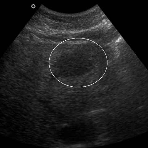
Patient history
A 67-year-old woman was referred for further assessment after a focal liver lesion was found on routine abdominal ultrasound. Physical examination revealed no symptoms or clinical findings. There was no history of weight loss, fever or other abnormality. Tumour markers were negative.
Imaging findings — key points
- Abdominal ultrasound shows a well-defined, oval, hypoechoic solid lesion in the liver with homogeneous echotexture and no posterior acoustic enhancement.
- Contrast-enhanced CT demonstrates a homogeneous, hypodense lesion in the hepatic parenchyma with mild, progressive enhancement and no central necrosis or calcification.
- MRI shows the lesion to be hypointense on T1-weighted images and mildly hyperintense on T2-weighted images, with restricted diffusion and homogeneous, mildly avid enhancement.
- Diffusion-weighted and perfusion imaging confirm a solid lesion with a low ADC value, without a capsule, internal septation or central scar.
- There is no hepatomegaly, lymphadenopathy, splenomegaly or other focal liver lesion, and the liver is not cirrhotic.
Discussion
Primary hepatic follicular lymphoma is a rare extranodal lymphoma that arises in the liver without systemic disease, and it usually presents as an incidental solitary mass in an otherwise healthy patient. Imaging shows a homogeneous solid lesion with mild enhancement, restricted diffusion and no central necrosis, but these features overlap with metastasis and other primary liver tumours, so biopsy is usually required. The main differential is secondary hepatic involvement by lymphoma, which is far more common and is accompanied by nodal or splenic disease, and metastatic disease. Prognosis is generally favourable, and treatment depends on histology and stage, ranging from observation to chemotherapy.
Differential diagnosis
| Alternative diagnosis | Key features that distinguish it from this case |
|---|---|
| Secondary hepatic lymphoma | Secondary lymphoma is accompanied by widespread lymphadenopathy, splenic involvement and systemic B symptoms, whereas this lesion is isolated to the liver without other disease. |
| Metastatic disease | Metastases are usually multiple, show ring enhancement and are associated with a known primary tumour, and they do not have the homogeneous mild enhancement of lymphoma. |
| Fungal microabscess | Fungal lesions are multiple, small, often with a target appearance and restricted diffusion, and occur in immunocompromised febrile patients. |
| Reactive lymphoid hyperplasia | Reactive lymphoid hyperplasia is a small benign nodule that is usually isointense to liver and does not show the restricted diffusion and progressive enhancement of lymphoma. |
The confirmed diagnosis itself is not included as an alternative — it is stated separately below.