Subungual epidermoid cyst
Written and reviewed by RadLearn editorial team · Updated 20 Oct 2026
Figures (16)
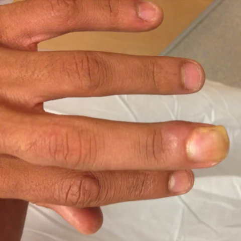
Patient history
A 30-year-old left-handed man presented with a subungual mass of the third finger of the left hand with secondary nail dystrophy (figure 1). The patient did not recall any injury to that finger.
Imaging findings — key points
- The clinical photograph shows a subungual mass with secondary nail dystrophy and ridging of the third finger of the left hand.
- The conventional radiograph shows soft-tissue swelling beneath the nail and a smooth pressure erosion of the dorsal cortex of the distal phalanx, without periosteal reaction or matrix calcification.
- Coronal and axial T1-weighted MRI show a well-defined subungual mass that is isointense to muscle and lifts the nail plate, with no invasion of the underlying bone marrow.
- The mass is hyperintense on proton density fat-saturated images and shows restricted diffusion, consistent with keratinaceous content, and it enhances peripherally without a solid enhancing nodule.
- There is no cortical destruction, marrow replacement or satellite lesion.
Discussion
Subungual epidermoid cyst is a rare lesion of the distal phalanx that may be congenital or follow penetrating trauma, and it presents as a slowly growing subungual mass with nail dystrophy and sometimes pain. Imaging shows a well-defined mass beneath the nail that erodes the dorsal cortex of the distal phalanx smoothly, with T2 hyperintensity and restricted diffusion from keratin, and peripheral rather than solid enhancement. The main differentials are malignant subungual tumours such as squamous cell carcinoma and keratoacanthoma, which show solid enhancement and bone destruction, and a glomus tumour. Treatment is surgical excision with nail bed repair, and the prognosis is excellent.
Differential diagnosis
| Alternative diagnosis | Key features that distinguish it from this case |
|---|---|
| Keratoacanthoma | A keratoacanthoma is a rapidly growing solid epidermal tumour with a central keratin plug that enhances and may invade the nail bed, unlike a simple subungual cyst. |
| Squamous cell carcinoma | Squamous cell carcinoma shows an irregular solid mass with bone destruction and marrow invasion, and it does not have the well-defined cystic appearance with peripheral enhancement. |
| Fibroma | A fibroma is a solid, low-T2-signal mass that enhances homogeneously and does not show the fluid-like, diffusion-restricted keratin content of an epidermoid cyst. |
| Glomus tumour | A glomus tumour is a small, intensely enhancing, hypervascular lesion in the subungual region that is exquisitely painful and cold-sensitive, without cystic keratin signal. |
The confirmed diagnosis itself is not included as an alternative — it is stated separately below.