Fasciitis with ischaemic features
Written and reviewed by RadLearn editorial team · Updated 20 Oct 2026
Figures (7)
Patient history
We describe a 65-year-old man who presented to our department with an asymptomatic mass in the right axilla. On clinical history the patient reported excision of a lipoma at the same site two years earlier.
Imaging findings — key points
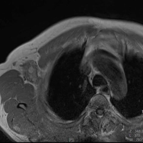
- MRI of the right axilla shows an ill-defined mass centred on the fascial planes between the subcutaneous fat and the axillary muscles, at the site of previous lipoma excision.
- The mass is isointense to muscle on T1-weighted images and hyperintense on T2-weighted images, with extensive surrounding oedema and thickening of the fascia.
- Dynamic contrast-enhanced T1-weighted images show heterogeneous enhancement of the peripheral solid component, with central non-enhancing areas consistent with ischaemic necrosis.
- There is no bone involvement, and the axillary vessels are patent without thrombosis.
- Pathology confirms a fasciitis with ischaemic features rather than a sarcoma.
Discussion
Fasciitis is a benign reactive proliferation of fibroblasts and myofibroblasts that arises from fascia and may follow local trauma or surgery, as in this case after lipoma excision. It usually presents as a rapidly growing mass and can mimic a sarcoma clinically and radiologically, and the presence of ischaemic features reflects outgrowth of the blood supply with central necrosis. MRI shows a fascial-based mass with surrounding oedema and heterogeneous enhancement, and the diagnosis is confirmed histologically. Recognition is important to avoid unnecessary radical surgery, and the lesion usually resolves with conservative management or simple excision.
Differential diagnosis
| Alternative diagnosis | Key features that distinguish it from this case |
|---|---|
| Soft-tissue sarcoma | A sarcoma is usually larger, with nodular enhancement, necrosis and invasion of adjacent structures, and it does not follow a fascial plane with the diffuse oedema typical of fasciitis. |
| Proliferative fasciitis | Proliferative fasciitis contains ganglion-like cells and is a histological variant, and it shows similar fascial oedema without the central ischaemic necrosis seen here. |
| Nodular fasciitis | Nodular fasciitis is a small, rapidly growing nodule with a fascial tail and homogeneous enhancement, rather than a large mass with central ischaemia. |
| Postoperative seroma or haematoma | A seroma or haematoma is a fluid collection at the surgical bed with a thin enhancing rim and no solid enhancing component. |
The confirmed diagnosis itself is not included as an alternative — it is stated separately below.