Spontaneous spinal epidural haematoma
Written and reviewed by RadLearn editorial team · Updated 20 Oct 2026
Figures (21)
Patient history
The patient developed severe, progressively worsening back pain and impaired gait over three days. After CT imaging, and given a recent history of infective spondylitis, a recurrent infective process was assumed and a conservative approach was adopted. On day 12 after admission, acute paraplegia developed.
Imaging findings — key points
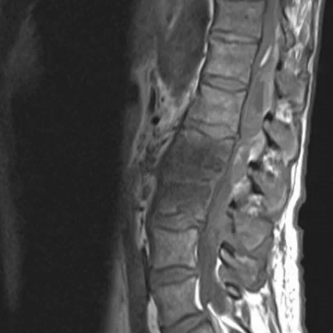
- Sagittal T1-weighted images of the lumbar and thoracic spine show a dorsal epidural mass extending over several vertebral levels, isointense to slightly hyperintense relative to the spinal cord, displacing the thecal sac anteriorly.
- The mass is heterogeneous on sagittal T2-weighted images, with areas of high signal indicating subacute haemorrhage, and it compresses the cord with effacement of the anterior and posterior cerebrospinal fluid spaces.
- Sagittal STIR images show hyperintensity within the epidural collection, confirming its fluid-haemorrhagic nature, while the adjacent vertebral bodies and discs remain normal in signal.
- Axial T2 fat-saturated images demonstrate a biconvex dorsal epidural collection narrowing the spinal canal and flattening the cord, without a paraspinal soft-tissue mass.
- Diffusion-weighted imaging and the ADC map show no restricted diffusion within the collection, and there is no peripheral enhancement, features that argue against an epidural abscess.
Discussion
Spontaneous spinal epidural haematoma is a rare cause of acute myelopathy produced by bleeding from the epidural venous plexus, often in the setting of anticoagulation, hypertension or a vascular malformation, and occasionally after minor trauma. The classic presentation is sudden severe back or neck pain followed by progressive paraparesis, and the rate of neurological decline determines the urgency of decompression. MRI is the investigation of choice: the haematoma is a biconvex dorsal epidural collection crossing multiple segments, with signal depending on the age of blood, and it must be separated from epidural abscess and metastasis, since the management and prognosis differ. Delay in diagnosis can convert a reversible compressive lesion into permanent cord infarction.
Differential diagnosis
| Alternative diagnosis | Key features that distinguish it from this case |
|---|---|
| Spinal epidural abscess | An epidural abscess usually presents with fever, raised inflammatory markers and vertebral discitis or osteomyelitis, and shows peripheral enhancement with restricted diffusion of pus. |
| Epidural metastasis | Metastatic epidural disease is accompanied by bone destruction, abnormal marrow signal and enhancement of the deposit, and often involves the vertebral body rather than forming a purely dorsal biconvex collection. |
| Epidural lipomatosis | Epidural lipomatosis follows the signal of fat on all sequences and suppresses on fat-saturated images, whereas a haematoma shows blood signal and STIR hyperintensity. |
| Disc herniation | A disc herniation is centred on an intervertebral disc level and is continuous with the disc, while a spontaneous epidural haematoma extends across several segments dorsal to the thecal sac. |
The confirmed diagnosis itself is not included as an alternative — it is stated separately below.