Metronidazole-induced encephalopathy
Written and reviewed by RadLearn editorial team · Updated 20 Oct 2026
Figures (20)
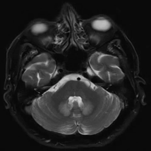
Patient history
A 59-year-old man treated with metronidazole for hidradenitis suppurativa over the previous two weeks presented to our clinic with several falls in recent days. Neurological examination showed cerebellar ataxia and mild dysarthria.
Imaging findings — key points
- Axial T2-weighted and FLAIR images show bilateral, symmetric hyperintensity in the cerebellar dentate nuclei, the dorsal pons and the medulla, with a characteristic involvement of the dentate nuclei.
- Additional hyperintensity is present in the splenium of the corpus callosum and in the periventricular white matter, without mass effect or surrounding oedema.
- Diffusion-weighted imaging shows restricted diffusion in the splenium and in the dentate nuclei, with corresponding low signal on the ADC map.
- No contrast enhancement, haemorrhage or abnormal meningeal enhancement is identified.
- Follow-up FLAIR and diffusion-weighted MRI five days after stopping metronidazole shows clear regression of the hyperintense lesions, supporting a reversible toxic aetiology.
Discussion
Metronidazole-induced encephalopathy is a reversible toxic encephalopathy that occurs after prolonged or high-dose metronidazole therapy and probably reflects axonal injury and intramyelinic oedema. The most characteristic imaging finding is bilateral symmetric hyperintensity of the cerebellar dentate nuclei on T2-weighted and FLAIR images, with involvement of the dorsal pons, medulla, splenium and periventricular white matter, and restricted diffusion may be present. The distribution differs from that of Wernicke encephalopathy, which involves the medial thalami and mammillary bodies, and from osmotic demyelination. Stopping the drug usually leads to clinical and radiological resolution within days to weeks, so recognition prevents unnecessary investigation and treatment.
Differential diagnosis
| Alternative diagnosis | Key features that distinguish it from this case |
|---|---|
| Acute Wernicke encephalopathy | Wernicke encephalopathy involves the medial thalami, periaqueductal grey matter and mammillary bodies and is associated with alcohol misuse or malnutrition, whereas metronidazole toxicity favours the dentate nuclei and splenium. |
| Enterovirus encephalomyelitis | Enterovirus infection produces a febrile illness with rhombencephalitis and often anterior horn cell involvement, and the lesions do not resolve simply on withdrawal of a drug. |
| Methyl bromide poisoning | Methyl bromide poisoning causes cerebellar and brainstem lesions after occupational exposure, and the history of exposure rather than drug intake is the discriminating factor. |
| Marchiafava-Bignami disease | Marchiafava-Bignami disease affects the corpus callosum in patients with chronic alcohol misuse and does not typically involve the dentate nuclei symmetrically. |
The confirmed diagnosis itself is not included as an alternative — it is stated separately below.