Colloid cyst of the third ventricle
Written and reviewed by RadLearn editorial team · Updated 20 Oct 2026
Figures (18)
Patient history
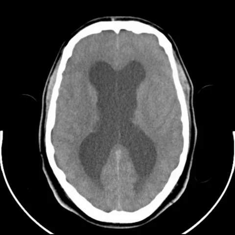
A 29-year-old man presented with a sudden, severe headache.
Imaging findings — key points
- Unenhanced CT demonstrates a well-defined, round, spontaneously hyperdense lesion at the foramen of Monro, measuring approximately 1 to 2 cm, without calcification and without surrounding oedema.
- There is dilatation of both lateral ventricles with a normal fourth ventricle, indicating obstruction at the level of the foramen of Monro.
- Cerebral digital subtraction angiography shows no tumour blush or pathological vascularity, confirming the avascular nature of the lesion.
- On MRI the lesion is hyperintense on T1-weighted images and iso- to hyperintense on T2-weighted images with a thin hypointense rim, and it does not enhance after gadolinium.
- Follow-up CT and MRI show a stable lesion with persistent ventricular dilatation but no interval growth or new hydrocephalus-related complication.
Discussion
Colloid cyst of the third ventricle is a benign epithelial-lined cyst filled with mucinous material that typically arises at the anterior roof of the third ventricle near the foramen of Monro. Its proteinaceous content makes it hyperdense on unenhanced CT and often hyperintense on T1-weighted MRI, and the absence of enhancement and of restricted diffusion helps to separate it from other third ventricular masses. The main clinical risk is acute obstructive hydrocephalus, which may cause sudden severe headache, collapse or death, so even an incidental cyst requires follow-up and consideration of resection when it is large or causes ventricular dilatation. Recognising the characteristic location and density avoids misinterpreting the lesion as a vascular or neoplastic mass.
Differential diagnosis
| Alternative diagnosis | Key features that distinguish it from this case |
|---|---|
| Choroid plexus cyst | A choroid plexus cyst follows cerebrospinal fluid signal on all sequences, is not hyperdense on unenhanced CT and does not contain the proteinaceous content responsible for T1 hyperintensity. |
| Craniopharyngioma | A craniopharyngioma arises in the suprasellar cistern, commonly contains calcification and cystic components and enhances, whereas a colloid cyst sits at the foramen of Monro without enhancement. |
| Neurocysticercosis | A cysticercal cyst shows a mural scolex, may have surrounding oedema in the vesicular stage and does not present as a hyperdense T1-hyperintense lesion at the foramen of Monro. |
| Forniceal or third ventricular glioma | A glioma is usually T2 hyperintense and enhances to a variable degree, and it tends to infiltrate rather than form a sharply marginated hyperdense cyst. |
The confirmed diagnosis itself is not included as an alternative — it is stated separately below.