Spontaneous intracranial hypotension
Written and reviewed by RadLearn editorial team · Updated 20 Oct 2026
Figures (16)
Patient history
A 37-year-old man had a one-month history of headache, worsening over the past week. The headache was reportedly worst on standing and partially relieved when supine. There was no history of fever, neck stiffness, trauma or spinal symptoms.
Imaging findings — key points
- T2-weighted, FLAIR and T1-weighted MRI show diffuse, smooth thickening and enhancement of the pachymeninges over the convexities, falx and tentorium, without leptomeningeal involvement.
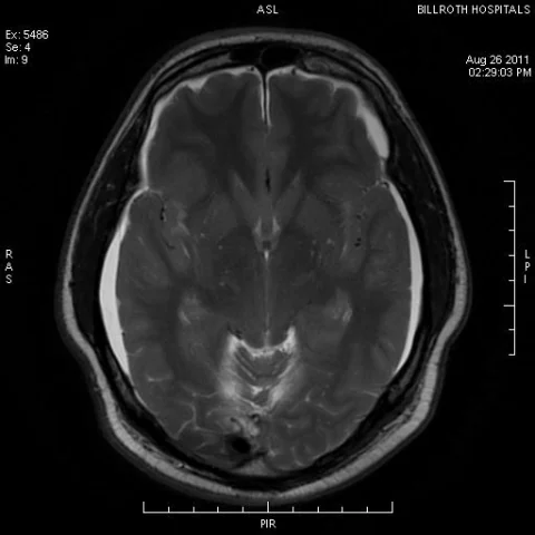
- There is sagging of the brainstem with brainstem descent, tonsillar descent and flattening of the pons against the clivus, together with reduced distance between the mammillary bodies and the pons.
- The dural venous sinuses and pituitary gland are engorged, and bilateral thin subdural effusions are present.
- Sagittal and axial T2-weighted images of the spine show an extensive dorsal epidural collection of cerebrospinal fluid-like signal extending over multiple levels, consistent with a spinal cerebrospinal fluid leak.
- No focal intraparenchymal lesion, subarachnoid haemorrhage or leptomeningeal enhancement is identified.
Discussion
Spontaneous intracranial hypotension results from a spontaneous cerebrospinal fluid leak, usually at the thoracic or cervical spine, and produces a low-pressure syndrome with orthostatic headache as its hallmark. Loss of cerebrospinal fluid volume causes compensatory venous engorgement and diffuse pachymeningeal enhancement, and the brain sags downward, which can mimic a Chiari malformation or a subdural collection. Spinal MRI is important to localise the leak, showing an extradural collection of cerebrospinal fluid, meningeal diverticula or a direct leak, and an epidural blood patch is the mainstay of treatment when conservative measures fail. The condition is under-recognised and can be mistaken for meningitis or a primary dural disease.
Differential diagnosis
| Alternative diagnosis | Key features that distinguish it from this case |
|---|---|
| Idiopathic hypertrophic pachymeningitis | Hypertrophic pachymeningitis produces nodular or mass-like dural thickening that may enhance intensely and does not cause brain sagging, venous engorgement or a spinal fluid collection. |
| Chronic subdural haematoma | A chronic subdural haematoma is a crescentic collection with blood signal and a membrane, often unilateral, without diffuse pachymeningeal enhancement or brainstem sagging. |
| Infective meningitis | Infective meningitis enhances the leptomeninges rather than the dura and is accompanied by fever, neck stiffness and cerebrospinal fluid pleocytosis. |
| Post-lumbar puncture headache | A post-lumbar puncture leak produces identical imaging findings but is distinguished by the procedure history, whereas spontaneous intracranial hypotension occurs without an obvious cause. |
The confirmed diagnosis itself is not included as an alternative — it is stated separately below.