Spigelian hernia
Written and reviewed by RadLearn editorial team · Updated 20 Oct 2026
Figures (7)
Patient history
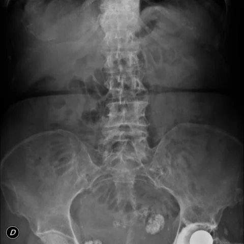
An elderly lady was admitted with colicky abdominal pain associated with nausea. Clinical examination and laboratory tests were unremarkable. Ultrasound and an abdominal radiograph were requested.
Imaging findings — key points
- The abdominal radiograph is unremarkable, with a normal bowel gas pattern and no evidence of obstruction.
- Ultrasound demonstrates a focal defect in the linea semilunaris with a small amount of herniated fat and fluid at the lateral border of the rectus abdominis.
- Multidetector CT confirms a Spigelian hernia through the aponeurosis of the transversus abdominis, lateral to the rectus muscle and below the level of the umbilicus.
- The hernia sac contains fat and a short segment of small bowel, with a narrow neck but no signs of strangulation or obstruction.
- There is no abdominal wall lipoma, haematoma or other hernia defect on the available images.
Discussion
Spigelian hernia occurs through the spigelian fascia, the aponeurotic layer between the rectus abdominis and the semilunar line, usually at or below the arcuate line where the posterior rectus sheath is deficient. It is often small and difficult to palpate, and patients present with intermittent abdominal pain or a palpable lump, with a risk of incarceration because of the narrow neck. CT is the most reliable investigation, showing the fascial defect and its contents, while ultrasound can be used in thin patients. Surgical repair is recommended even for asymptomatic hernias because of the high risk of strangulation.
Differential diagnosis
| Alternative diagnosis | Key features that distinguish it from this case |
|---|---|
| Other abdominal wall hernia | Inguinal, femoral and ventral hernias occur at their own anatomic landmarks and are not located lateral to the rectus muscle at or below the arcuate line. |
| Abdominal wall lipoma | A lipoma is a homogeneous fat-density mass within the subcutaneous tissue or muscle, without a fascial defect or a neck connecting it to the peritoneal cavity. |
| Rectus sheath haematoma | A rectus sheath haematoma is a fluid or blood-density collection within the rectus sheath, often after anticoagulation or trauma, without a fascial defect. |
| Spigelian hernia with strangulation | Strangulation would show bowel wall thickening, reduced enhancement, fluid in the sac and obstruction, none of which are present in this case. |
The confirmed diagnosis itself is not included as an alternative — it is stated separately below.