Coeliac artery compression by the median arcuate ligament
Written and reviewed by RadLearn editorial team · Updated 20 Oct 2026
Figures (7)
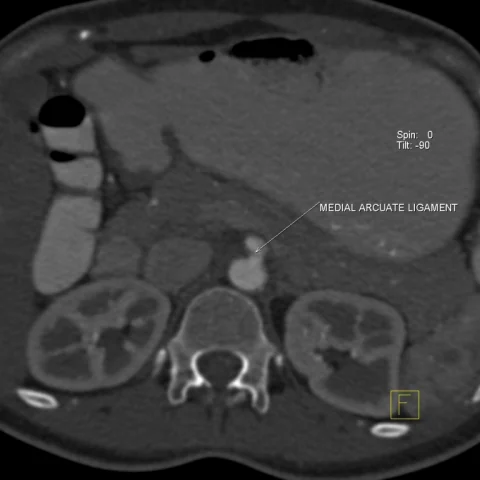
Patient history
A 28-year-old woman had severe epigastric pain immediately after meals, lasting one hour, over the past six months. Nothing relieved her pain. On examination she appeared unwell. Ultrasound showed no significant findings. She was then referred for contrast-enhanced abdominal CT.
Imaging findings — key points
- Axial contrast-enhanced abdominal CT is normal, and sagittal thin-slice MDCT reconstruction demonstrates a focal, hook-like compression of the proximal coeliac trunk by the median arcuate ligament.
- The stenosis is eccentric, with post-stenotic dilatation of the coeliac artery and a characteristic inferior indentation of the vessel.
- Doppler ultrasound shows a markedly increased peak systolic velocity in the coeliac artery, exceeding 200 cm/s, with turbulent flow at the stenosis.
- The velocity and degree of narrowing increase during expiration and decrease during inspiration, confirming dynamic compression by the ligament.
- No atherosclerosis, thrombus, aneurysm or extrinsic mass is present in the coeliac artery or the superior mesenteric artery.
Discussion
Median arcuate ligament syndrome, also called coeliac artery compression syndrome, results from compression of the coeliac trunk by the median arcuate ligament of the diaphragm, and it is a diagnosis of exclusion because the same imaging finding occurs in many asymptomatic people. The classic patient is a young woman with postprandial epigastric pain, weight loss and sometimes an abdominal bruit, and the narrowing characteristically worsens in expiration. CT angiography with sagittal reformats and Doppler ultrasound showing elevated peak systolic velocity are the key investigations, and other causes of abdominal pain must be excluded. Treatment options include laparoscopic release of the ligament and, in selected cases, revascularisation.
Differential diagnosis
| Alternative diagnosis | Key features that distinguish it from this case |
|---|---|
| Superior mesenteric artery thrombosis | SMA thrombosis produces an occluded or severely narrowed artery with bowel ischaemia and a non-enhancing bowel wall, rather than extrinsic compression of the coeliac trunk. |
| Abdominal malignancy | A retroperitoneal or pancreatic malignancy encases or invades the vessel with soft-tissue mass, lymphadenopathy and weight loss, and does not produce a smooth hook-like indentation. |
| Atherosclerotic coeliac stenosis | Atherosclerotic stenosis occurs in older patients with calcified plaque at the vessel origin and is fixed, without the expiratory accentuation of median arcuate ligament compression. |
| Functional abdominal pain | Functional abdominal pain has no vascular abnormality, whereas this case shows a reproducible anatomic narrowing with elevated velocities and post-stenotic dilatation. |
The confirmed diagnosis itself is not included as an alternative — it is stated separately below.