Small bowel diverticulosis
Written and reviewed by RadLearn editorial team · Updated 20 Oct 2026
Figures (5)
Patient history
A 73-year-old man had a history of intermittent abdominal discomfort and bloating or fullness dating back several years. Laboratory tests requested by his GP were unremarkable apart from iron-deficiency anaemia. The patient declined endoscopy.
Imaging findings — key points
- The abdominal radiograph is non-specific, showing a non-obstructed bowel gas pattern with no pneumoperitoneum or dilated loops.
- Contrast-enhanced CT demonstrates multiple gas- and fluid-filled outpouchings arising from the mesenteric border of the jejunum and ileum, with thin walls and no surrounding inflammatory change.
- The diverticula vary in size and some contain ingested material or small air-fluid levels, and the adjacent small bowel wall is normal in thickness.
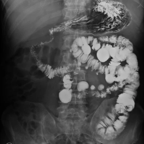
- A barium meal confirms numerous contrast-filled diverticula along the small bowel, without stricture, mass or obstruction.
- There is no evidence of diverticulitis, perforation, abscess or mesenteric venous thrombosis.
Discussion
Small bowel diverticulosis is an acquired condition in which mucosa herniates through the muscular layer, usually along the mesenteric border where the vasa recta penetrate, and it is most common in the jejunum. Most patients are asymptomatic or have vague abdominal discomfort, but complications include diverticulitis, perforation, obstruction, haemorrhage and bacterial overgrowth with malabsorption. CT shows multiple thin-walled outpouchings with gas, fluid or ingested material, and the diagnosis is confirmed on barium studies. Uncomplicated diverticulosis requires no treatment, while complications are managed supportively or surgically depending on severity.
Differential diagnosis
| Alternative diagnosis | Key features that distinguish it from this case |
|---|---|
| Small bowel neoplasm | A small bowel neoplasm produces a focal mass or wall thickening that enhances and may obstruct or bleed, without multiple thin-walled outpouchings along the mesenteric border. |
| Meckel diverticulum | A Meckel diverticulum is a single outpouching on the antimesenteric border of the distal ileum, not multiple diverticula distributed along the small bowel. |
| Diverticulitis | Diverticulitis shows wall thickening, fat stranding, fluid and possibly an abscess around the diverticula, whereas uncomplicated diverticulosis has no inflammatory change. |
| Pneumatosis intestinalis | Pneumatosis intestinalis is gas within the bowel wall itself, often linear or cystic, and is not a set of epithelial-lined outpouchings. |
The confirmed diagnosis itself is not included as an alternative — it is stated separately below.