Silicone-induced breast implant capsule granuloma (SIGBIC)
Written and reviewed by RadLearn editorial team · Updated 20 Oct 2026
Figures (12)
Patient history
In 2006, a 50-year-old woman with a BRCA1 mutation and a history of right breast cancer underwent right mastectomy with subpectoral reconstruction. The implant was a silicone gel implant with a pure polyurethane surface, elliptical base, and anatomical contour with an inferior pole. She attended our breast unit for annual follow-up. Clinical examination revealed mild breast asymmetry, left-dominant, but no palpable lump, nipple discharge, or local pain.
Imaging findings — key points
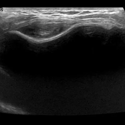
- Ultrasound shows two circumscribed oval intracapsular masses with a heterogeneous echopattern between the implant and the fibrous capsule in the upper outer and lower outer quadrants, with no sign of implant rupture.
- MRI confirms an intact implant without intra- or extracapsular rupture.
- In the lower outer quadrant, a 3 cm oval solid mass within the fibrous capsule is displaced medially against the implant.
- The mass shows heterogeneous signal on T2-weighted images, a central focus of restricted diffusion on the ADC map and progressive enhancement after gadolinium administration.
- A smaller lesion with identical MRI features is present in the upper outer quadrant.
- A thin layer of free fluid surrounds the lesions and the implant.
Discussion
Silicone-induced granuloma of the breast implant capsule (SIGBIC) results from silicone microdroplets that bleed through the elastomer of an otherwise intact implant and trigger an inflammatory foreign-body giant-cell reaction with collagen deposition in the fibrous capsule; it is usually an incidental finding but may cause capsular contracture. On ultrasound it appears as an intracapsular heterogeneous mass that may resemble a storm of debris, and MRI characteristically shows a T2-heterogeneous, T1-hypointense intracapsular mass with progressive (type I) enhancement, sometimes with the black drop sign. Because it can mimic BIA-ALCL, which typically presents with a large non-resolving seroma, the imaging findings must be interpreted carefully, and surgical removal with histopathology was performed here to confirm the diagnosis.
Differential diagnosis
| Alternative diagnosis | Key features that distinguish it from this case |
|---|---|
| Breast implant-associated anaplastic large cell lymphoma (BIA-ALCL) | BIA-ALCL usually presents as a large peri-implant seroma that does not resolve spontaneously and is often associated with capsular contracture, but it lacks the well-circumscribed, progressively enhancing solid intracapsular nodule seen here. |
| Late seroma | A late seroma is a simple fluid collection without a solid enhancing component or nodular areas, whereas this lesion contains a solid intracapsular mass. |
| Intracapsular haematoma | An intracapsular haematoma shows signal intensity that varies with the age of blood and no progressive nodular enhancement, unlike the enhancing solid mass here. |
| Breast carcinoma | Primary breast carcinoma arises in the glandular tissue rather than within the implant capsule and shows spiculated morphology with washout kinetics, not a circumscribed intracapsular mass. |
The confirmed diagnosis itself is not included as an alternative — it is stated separately below.