Idiopathic granulomatous mastitis
Written and reviewed by RadLearn editorial team · Updated 20 Oct 2026
Figures (11)
Patient history
A 27-year-old woman presented with a gradually enlarging mass in the subareolar region of the left breast, accompanied by swelling and mastalgia for one week. She had been breastfeeding for two years and had weaned one year prior.
Imaging findings — key points
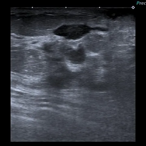
- Ultrasound demonstrates an ill-defined, irregular, heterogeneous hypoechoic mass in the subareolar region of the left breast, composed of confluent tubular and nodular components.
- Within the mass there are small anechoic foci corresponding to microabscesses, and power Doppler shows increased peripheral and septal vascularity.
- The overlying skin and subcutaneous fat are thickened and oedematous, and there is no posterior acoustic shadowing of the kind usually produced by a scirrhous carcinoma.
- Follow-up ultrasound one month later shows progression of the inflammatory collection with additional microabscesses and early sinus tract formation.
- MRI shows an ill-defined regional area of non-mass enhancement with peripheral rim-like enhancement of the microabscesses, surrounded by T2 hyperintense oedema.
Discussion
Idiopathic granulomatous mastitis is a benign chronic inflammatory disease of the breast that typically affects young women, often within a few years of childbirth and breastfeeding. Imaging shows an ill-defined inflammatory mass with microabscesses in a subareolar location, and histology is essential because non-caseating granulomatous lobulitis with negative cultures both establishes the diagnosis and excludes infection and malignancy. Treatment is usually with corticosteroids or immunosuppression, and relapse after reduction of therapy, as occurred in this patient, is well recognised.
Differential diagnosis
| Alternative diagnosis | Key features that distinguish it from this case |
|---|---|
| Breast tuberculosis | Usually presents as a cold abscess with chronic sinus tracts and caseating granulomas, with acid-fast bacilli or a positive culture or PCR result, and is often accompanied by necrotic axillary nodes with calcification. |
| granulomatosis with polyangiitis | Produces granulomatous inflammation with vasculitis accompanied by upper-airway, pulmonary or renal disease and positive ANCA serology. |
| Carcinoma (inflammatory or medullary) | Shows a discrete solid mass with suspicious microcalcifications, spiculated margins and axillary adenopathy, and does not resolve with corticosteroid therapy. |
The confirmed diagnosis itself is not included as an alternative — it is stated separately below.