Primary breast lymphoma, diffuse large B-cell
Written and reviewed by RadLearn editorial team · Updated 20 Oct 2026
Figures (10)
Patient history
A 63-year-old woman was referred to the breast clinic due to a 3-month history of a painless, palpable breast mass. The patient had a history of surgical excision of benign breast lesions. Clinical examination revealed two nodules in the right breast, with no skin or nipple changes. No axillary lymphadenopathy was detected.
Imaging findings — key points
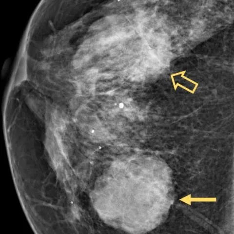
- Mammography shows two high-density masses at the junction of the right inner quadrant and the upper outer quadrant, one with smooth margins and the other with partially obscured margins.
- Ultrasound demonstrates two heterogeneous masses: a round, indistinctly marginated, non-parallel lesion with posterior acoustic enhancement and internal vascularity, and a second oval, circumscribed lesion with parallel orientation and no posterior acoustic feature.
- Axial T2-weighted MRI shows two round to oval masses of intermediate signal with circumscribed and lobulated margins, associated with mild distortion of the surrounding parenchyma.
- Dynamic contrast-enhanced MRI shows rapid heterogeneous enhancement with predominantly peripheral washout on the maximum intensity projection image.
- DWI demonstrates true diffusion restriction with markedly low ADC values of 0.57-0.58 mm2/s.
- FDG-PET/CT shows increased FDG uptake confined to the breast lesions, with no other abnormal site of uptake.
Discussion
Primary breast lymphoma is a rare haematological malignancy accounting for less than 1% of breast malignancies, with diffuse large B-cell lymphoma the most common subtype and a median patient age of 60-65 years. Imaging features are non-specific and overlap with breast carcinoma, but a circumscribed round or oval hyperdense mass without calcification, hypervascularity on Doppler, heterogeneous enhancement with washout and markedly reduced ADC values should raise suspicion of lymphoma. Diagnosis requires core needle biopsy with histopathological and immunohistochemical confirmation, and FDG-PET/CT is regarded as the most accurate technique for staging FDG-avid lymphoma and assessing treatment response. Treatment is primarily chemotherapy, as surgical resection has shown no additional benefit.
Differential diagnosis
| Alternative diagnosis | Key features that distinguish it from this case |
|---|---|
| Invasive carcinoma of no special type | Usually shows spiculated or irregular margins with architectural distortion and possible microcalcification, whereas these lesions are round, circumscribed and non-calcified with very low ADC values favouring lymphoma. |
| Fibroadenoma | A circumscribed oval hypoechoic mass parallel to the skin with uniform internal echoes and no diffusion restriction, unlike the heterogeneous washout enhancement and profound ADC reduction seen here. |
| Phyllodes tumour | A large circumscribed lobulated mass with cystic spaces and rapid growth, lacking the homogeneous intermediate T2 signal and marked diffusion restriction of this lesion. |
| Medullary carcinoma | A well-circumscribed soft round mass with a pushing border and prominent lymphoplasmacytic infiltrate, but it does not show the extremely low ADC values produced by tightly packed lymphoma cells. |
| Breast metastasis | Presents as multiple bilateral round circumscribed nodules in a patient with a known extramammary primary; the absence of such a history and the unilateral dominant presentation favour a primary breast lymphoma. |
The confirmed diagnosis itself is not included as an alternative — it is stated separately below.