Recurrent ameloblastoma of the right jaw after conservative surgery
Written and reviewed by RadLearn editorial team · Updated 20 Oct 2026
Figures (18)
Patient history
A 40-year-old woman recently presented to our department with bleeding from the right lower dental arch, gingival pain and difficulty moving the tongue.
Imaging findings — key points
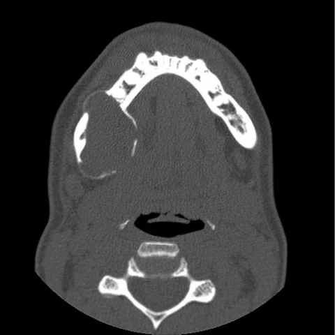
- CT shows an expansile, multilocular, predominantly cystic lesion of the right posterior mandible with thinned and focally perforated cortices and resorption of the roots of the adjacent molar teeth.
- The lesion extends beyond the previous surgical site into the surrounding soft tissues of the masticator and sublingual spaces, indicating recurrence.
- MRI shows a mixed solid and cystic mass with hyperintense locules on T2-weighted images and intermediate-signal solid components that enhance after gadolinium.
- Follow-up CT and MRI demonstrate progressive enlargement of the lesion with new cortical breakthrough and displacement of the inferior alveolar canal.
- There is no internal calcification or ossification, and no enlarged cervical lymph nodes are identified.
Discussion
Ameloblastoma is a benign but locally aggressive odontogenic tumour that arises from the enamel organ or dental lamina and most commonly affects the posterior mandible in the fourth to fifth decade. Conservative treatment by enucleation or curettage is followed by recurrence in a substantial proportion of cases, particularly when the tumour has broken through cortex or involved the surrounding soft tissue, which is why long-term imaging follow-up is mandatory. CT defines the extent of bone destruction and cortical breakthrough, while MRI is superior for detecting soft-tissue extension and solid enhancing components that predict a higher risk of recurrence. Radical resection with clear margins is the treatment of choice for recurrent disease.
Differential diagnosis
| Alternative diagnosis | Key features that distinguish it from this case |
|---|---|
| Odontogenic keratocyst | An odontogenic keratocyst is usually unilocular, grows along the bone without marked expansion, does not resorb tooth roots and shows restricted diffusion from its keratin content. |
| Central giant cell granuloma | Central giant cell granuloma typically affects the anterior mandible of young patients, is multilocular with wispy septa and does not show the solid enhancing components of an ameloblastoma. |
| Calcifying epithelial odontogenic tumour | A calcifying epithelial odontogenic tumour contains scattered calcifications and amyloid-like material and is often associated with an impacted tooth. |
| Odontogenic myxoma | Odontogenic myxoma produces a fine, soap-bubble trabecular pattern and high T2 signal from myxoid stroma, without the solid enhancing nodules seen here. |
| Ossifying fibroma | Ossifying fibroma has a well-defined bony shell with internal ossification that progresses from ground-glass to dense, unlike the purely lytic recurrent lesion described. |
The confirmed diagnosis itself is not included as an alternative — it is stated separately below.