Desmoplastic fibroma of the mandible
Written and reviewed by RadLearn editorial team · Updated 20 Oct 2026
Figures (12)
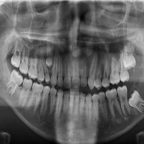
Patient history
A 15-year-old boy presented with pain and a palpable mass in the left facial region. He reported a three-month history and limited mouth opening. There was no fever and no history of recent trauma. On examination the mass was hard.
Imaging findings — key points
- Frontal radiography and axial CT show a large, expansile, multilocular lytic lesion of the left mandible with well-defined but thinned cortices and no internal calcification or ossification.
- The lesion expands the buccal and lingual cortex and extends into the adjacent soft tissues of the masticator space, and several tooth roots are displaced.
- Axial T1-weighted MRI shows the lesion to be isointense to muscle, and T2-weighted images show predominantly low to intermediate signal with heterogeneous hyperintense areas, reflecting dense fibrous tissue.
- Coronal STIR images show mild surrounding oedema, and diffusion-weighted imaging shows no marked restricted diffusion.
- Axial VIBE and coronal T1-weighted images after contrast show heterogeneous, moderate enhancement of the solid components, and postoperative volume-rendered CT documents the surgical defect.
Discussion
Desmoplastic fibroma is a rare benign but locally aggressive fibromatous tumour of bone that most often affects the mandible, femur or pelvis in children and young adults. Histologically it resembles desmoid fibromatosis, and its dense collagen content explains the low-to-intermediate T2 signal that helps to distinguish it from other lytic bone lesions. It grows slowly and produces pain, swelling and limited mouth opening, but it can erode cortex and extend into soft tissue, and recurrence is common after curettage. Wide surgical excision is therefore preferred, and imaging follow-up is required because the tumour may recur many years later.
Differential diagnosis
| Alternative diagnosis | Key features that distinguish it from this case |
|---|---|
| Fibromatosis | Desmoid-type fibromatosis is a soft-tissue proliferation that infiltrates muscle and fascia without a well-defined osseous shell, whereas desmoplastic fibroma is centred within bone and expands the cortex. |
| Rhabdomyosarcoma | Rhabdomyosarcoma in a teenager is aggressive, destroys bone, shows marked enhancement and restricted diffusion and grows rapidly, in contrast with the slow, expansile growth of a desmoplastic fibroma. |
| Neuroblastoma metastasis | Neuroblastoma metastasis produces permeative bone destruction with periosteal reaction and often calcification, and is associated with an elevated urinary catecholamine level in a younger child. |
| Lymphoma of bone | Primary bone lymphoma causes permeative destruction with a large soft-tissue component, homogeneous enhancement and restricted diffusion, without the fibrous, low-T2 matrix of desmoplastic fibroma. |
The confirmed diagnosis itself is not included as an alternative — it is stated separately below.