Giant pontine cavernoma
Written and reviewed by RadLearn editorial team · Updated 20 Oct 2026
Figures (17)
Patient history
A 15-year-old girl was brought to our hospital with a Glasgow Coma Scale score consistent with coma. Her parents reported headache over the previous two days. No other previous medical problems were reported.
Imaging findings — key points
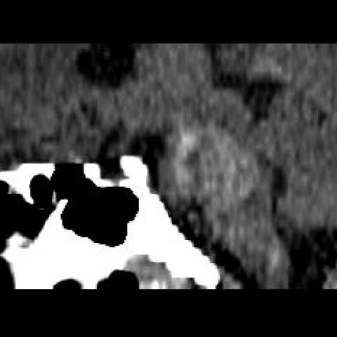
- Unenhanced CT shows a large hyperdense mass in the pons with surrounding low attenuation, indicating acute haemorrhage with oedema and mass effect on the fourth ventricle.
- T2* gradient-echo images demonstrate marked blooming artefact within the lesion, consistent with haemosiderin deposition from repeated haemorrhage.
- Axial T2-weighted images show a heterogeneous, multilobulated lesion with a popcorn-like core of mixed high and low signal and a complete hypointense haemosiderin rim.
- Pre-contrast T1-weighted images show areas of intrinsic high signal from subacute blood, with little or no enhancement after gadolinium administration.
- Sagittal and coronal T2 dark-fluid images confirm expansion of the pons with compression of the fourth ventricle and obstructive hydrocephalus, and postoperative CT shows a decompressed posterior fossa.
Discussion
Cavernous malformation is a low-flow vascular lesion composed of dilated sinusoidal channels without intervening brain parenchyma, and repeated small haemorrhages produce a characteristic popcorn-like core with a haemosiderin rim. Brainstem cavernomas are particularly dangerous because even a small bleed can cause catastrophic neurological deficit, and a giant lesion may produce coma from mass effect and hydrocephalus. MRI is diagnostic when T2* blooming and a complete haemosiderin rim are seen, but a giant lesion with recent haemorrhage requires exclusion of an underlying arteriovenous malformation or tumour. Management is individualised between conservative observation and microsurgical resection, depending on the number of bleeds, accessibility and neurological status.
Differential diagnosis
| Alternative diagnosis | Key features that distinguish it from this case |
|---|---|
| Diffuse pontine glioma | A diffuse pontine glioma expands the pons with T2 hyperintensity, lacks the popcorn appearance and haemosiderin rim, and does not show blooming on T2* imaging. |
| Arteriovenous malformation | An arteriovenous malformation contains flow voids and enlarged feeding arteries or draining veins, and haemorrhage is usually associated with these vascular structures. |
| Hypertensive haemorrhage | Hypertensive haemorrhage occurs typically in the basal ganglia, thalamus or pons in older patients with longstanding hypertension and shows a single acute haematoma rather than a multilobulated lesion with a haemosiderin rim. |
| Metastasis | A metastasis shows surrounding vasogenic oedema, usually enhances and is often multiple, without the complete haemosiderin rim of a cavernoma. |
The confirmed diagnosis itself is not included as an alternative — it is stated separately below.