Hydrocephalus
Written and reviewed by RadLearn editorial team · Updated 20 Oct 2026
Figures (19)
Patient history
A term infant with hydrocephalus, hepatomegaly, micropenis and hydronephrosis.
Imaging findings — key points
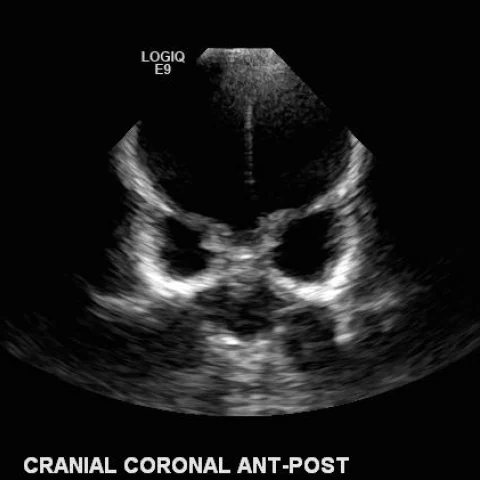
- Transcranial ultrasound through the anterior fontanelle shows marked dilatation of both lateral ventricles with a dangling choroid plexus and thinning of the cortical mantle.
- Unenhanced CT confirms severe ventriculomegaly involving the lateral and third ventricles, with temporal horn dilatation and no obstructing mass at the foramen of Monro.
- Coronal CT reformats show a widened interhemispheric fissure and thinned cortex, and 3D volume-rendered images demonstrate calvarial expansion with separated sutures.
- Axial and sagittal T1-weighted MRI show the same ventricular dilatation and help to exclude a posterior fossa mass or aqueductal web as the cause.
- The liver is enlarged and both kidneys show pelvicalyceal dilatation, indicating that the intracranial findings are part of a multisystem disorder rather than isolated hydrocephalus.
Discussion
Hydrocephalus in a term infant may be congenital or acquired and results from imbalance between cerebrospinal fluid production and absorption, whether by obstruction, dysresorption or, rarely, overproduction. The combination of hydrocephalus with hepatomegaly, micropenis and hydronephrosis suggests a syndromic or metabolic cause, and imaging is directed at identifying the level of obstruction and any associated malformation. Ultrasound through the open fontanelle is the first-line test, with CT and MRI used to define ventricular anatomy, cortical thickness and the state of the posterior fossa. Early recognition and diversion of cerebrospinal fluid are important because sustained raised pressure compromises the developing brain.
Differential diagnosis
| Alternative diagnosis | Key features that distinguish it from this case |
|---|---|
| Alobar holoprosencephaly | Holoprosencephaly shows a single monoventricle with fused thalami and absent midline structures, whereas this case has two separate lateral ventricles with a preserved midline. |
| Porencephalic cyst | A porencephalic cyst is a focal parenchymal cavity communicating with the ventricle and surrounded by gliosis, not a uniform dilatation of the entire ventricular system. |
| Fetal hydrocephalus | Fetal hydrocephalus is diagnosed in utero and may resolve or progress after birth, whereas this infant has a term delivery with established postnatal ventriculomegaly and systemic anomalies. |
| Aqueductal stenosis | Aqueductal stenosis dilates the lateral and third ventricles with a normal fourth ventricle, and it produces a prominent upstream dilation rather than diffuse, symmetric ventriculomegaly. |
The confirmed diagnosis itself is not included as an alternative — it is stated separately below.