Pulmonary tuberculosis
Written and reviewed by RadLearn editorial team · Updated 20 Oct 2026
Figures (18)
Patient history
A 53-year-old African man was referred to our institution for radiotherapy of an inoperable oesophageal squamous cell carcinoma diagnosed three months earlier. When the diagnosis was confirmed by endoscopic biopsy he reported episodes of dysphagia and weight loss. Laboratory tests showed anaemia (Hb 9.6 g/L), hypoalbuminaemia and a raised CRP.
Imaging findings — key points
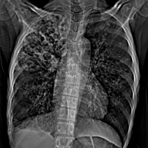
- The CT scout view shows upper-zone opacity, and the mediastinal window images demonstrate consolidation with cavitation in the upper lobes and enlarged mediastinal nodes.
- CT in the lung window shows a tree-in-bud pattern of centrilobular nodules in the upper lobes, together with areas of consolidation and a thick-walled cavity.
- HRCT confirms centrilobular nodules, branching linear opacities and subpleural consolidation, with architectural distortion and traction bronchiectasis in the affected lobes.
- There is a small pleural effusion and a calcified mediastinal lymph node, without significant lymphadenopathy elsewhere.
- Coronal reconstruction shows bilateral upper-lobe involvement, with relative sparing of the lower zones.
Discussion
Pulmonary tuberculosis remains an important diagnosis in patients with upper-lobe consolidation, cavitation and tree-in-bud nodularity, particularly in those with a relevant epidemiological background or immunosuppression, and it may coexist with malignancy. The tree-in-bud pattern reflects endobronchial spread of infection, while cavitation occurs when caseous necrosis communicates with a bronchus, and calcified lymph nodes suggest healed granulomatous disease. Imaging cannot reliably separate tuberculosis from other cavitating infections or a cavitating carcinoma, so microbiological confirmation is essential. Prolonged multidrug therapy is required, and the possibility of drug resistance should be considered in patients with an adequate treatment history.
Differential diagnosis
| Alternative diagnosis | Key features that distinguish it from this case |
|---|---|
| Atypical pneumonia | Atypical pneumonia usually shows lower-zone ground-glass opacity and consolidation that resolves with antibiotics, without tree-in-bud nodules, cavitation or calcified nodes. |
| Lung carcinoma | Lung carcinoma produces a dominant spiculated mass that enhances and may cavitate, but it does not give bilateral tree-in-bud nodularity or calcified lymph nodes. |
| Bronchiectasis | Bronchiectasis shows dilated, thick-walled airways with mucoid impaction, and it may coexist with tuberculosis but does not explain the cavitating consolidation and tree-in-bud pattern at presentation. |
| Aspergillosis | Invasive aspergillosis produces nodules with a halo sign or a cavitating lesion with an air crescent in immunosuppressed patients, whereas a mycetoma forms within a pre-existing cavity. |
The confirmed diagnosis itself is not included as an alternative — it is stated separately below.