Pulmonary nocardiosis in HIV
Written and reviewed by RadLearn editorial team · Updated 20 Oct 2026
Figures (23)
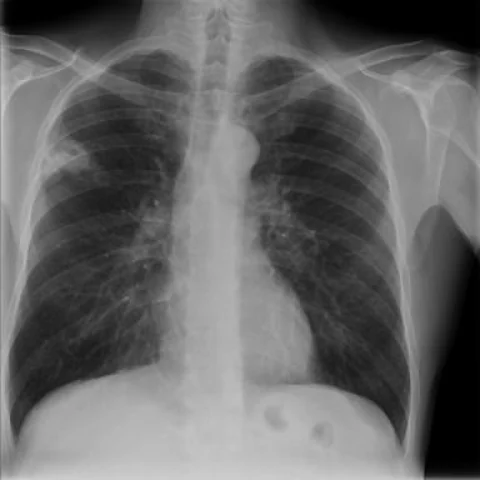
Patient history
A 45-year-old man with long-standing human immunodeficiency virus (HIV) infection and severe immunosuppression unresponsive to antiretroviral therapy (4 CD4+ cells/mm3) was admitted with malaise, diarrhoea and weight loss. He had a metabolic acidosis and no specific respiratory symptoms.
Imaging findings — key points
- The initial chest radiograph shows patchy consolidation in the right mid and lower zones with ill-defined nodular opacities, and early follow-up radiographs show progression despite antibiotic therapy.
- High-resolution CT demonstrates multiple bilateral nodules and masses of varying size, several of which cavitate, together with areas of consolidation and ground-glass opacity.
- The nodules have a random or clustered distribution without a tree-in-bud pattern, and there is no significant mediastinal lymphadenopathy.
- Subsequent HRCT shows coalescence of the lesions into larger cavitating masses with thick, irregular walls, and a small right pleural effusion.
- Post-treatment radiographs and HRCT show marked resolution of the consolidation and cavitation with residual scarring.
Discussion
Pulmonary nocardiosis is an opportunistic infection caused by Nocardia species that most often affects immunocompromised patients, particularly those with advanced HIV, long-term corticosteroid therapy or transplantation. The organism produces a suppurative necrotising pneumonia with a tendency to cavitate and to spread haematogenously to the brain and skin, and imaging shows multiple nodules, masses and consolidation that may mimic tuberculosis, fungal infection or malignancy. The diagnosis requires culture or molecular identification from respiratory samples or biopsy, because the imaging pattern is non-specific. Treatment is prolonged with trimethoprim-sulfamethoxazole or an alternative regimen, and the prognosis depends on early recognition and restoration of immunity.
Differential diagnosis
| Alternative diagnosis | Key features that distinguish it from this case |
|---|---|
| Tuberculosis | Pulmonary tuberculosis favours the upper lobes with tree-in-bud nodularity, cavitation and calcified lymph nodes, whereas nocardiosis produces multiple random nodules and masses without a tree-in-bud pattern. |
| Bacterial pneumonia | Ordinary bacterial pneumonia produces lobar or segmental consolidation that responds rapidly to antibiotics, rather than multiple cavitating nodules progressing over weeks. |
| Actinomycosis | Actinomycosis typically crosses tissue planes to involve the chest wall and pleura with sinus tracts, and it is not characterised by bilateral random nodules. |
| Septic embolism | Septic emboli are peripheral, wedge-shaped and cavitary with a feeding vessel, and they occur in patients with endocarditis or indwelling lines, often with positive blood cultures. |
| Cavitating metastasis | Cavitating metastases are usually multiple and peripheral in a patient with a known primary, and they do not resolve with antibiotic therapy alone. |
The confirmed diagnosis itself is not included as an alternative — it is stated separately below.