Endobronchial carcinoid
Written and reviewed by RadLearn editorial team · Updated 20 Oct 2026
Figures (12)
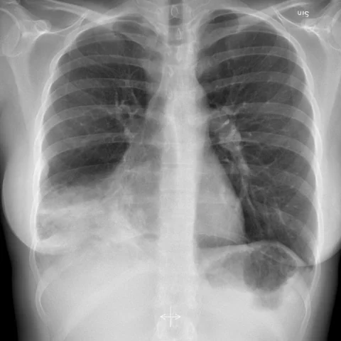
Patient history
A 37-year-old female non-smoker presented with recurrent pneumonia. She felt tired and had occasional chest pain, but no cough or dyspnoea. Auscultation of the lungs was otherwise unremarkable. Laboratory data included a slightly raised C-reactive protein and leukocytosis.
Imaging findings — key points
- The chest radiograph shows recurrent patchy consolidation and volume loss in the right lower lobe, with a rounded hilar opacity suggestive of a central obstructing lesion.
- CT and virtual bronchoscopy demonstrate a well-defined, rounded endobronchial nodule arising from the wall of a segmental bronchus, with a broad base and smooth surface, causing near-complete obstruction.
- Distal to the obstruction there is atelectasis with mucus-filled dilated bronchi producing a finger-in-glove appearance, and the lesion enhances avidly after contrast.
- PET-CT shows only low to moderate FDG uptake within the nodule, and the octreotide scan demonstrates somatostatin receptor expression, supporting a neuroendocrine tumour.
- Postoperative chest radiographs show re-expansion of the right lower lobe after resection.
Discussion
Endobronchial carcinoid is a low-grade neuroendocrine tumour that arises from Kulchitsky cells in the bronchial mucosa and most often occurs in the central airways of middle-aged non-smokers. It grows slowly as a polypoid endobronchial mass and causes recurrent pneumonia, atelectasis and haemoptysis, and the combination of a well-defined enhancing nodule, distal bronchocele and low FDG avidity with somatostatin receptor expression is characteristic. The main differentials are other endobronchial tumours and a foreign body granuloma, and biopsy is usually required for confirmation. Surgical resection with bronchoplastic reconstruction is curative in most patients, and the prognosis is excellent for typical carcinoid.
Differential diagnosis
| Alternative diagnosis | Key features that distinguish it from this case |
|---|---|
| Adenoid cystic carcinoma or mucoepidermoid carcinoma | These salivary-type tumours of the airway tend to have a broader base with submucosal infiltration, may destroy cartilage and show higher FDG uptake, rather than a smooth polypoid endobronchial nodule. |
| Foreign body granuloma | A foreign body granuloma occurs after aspiration, contains a radiopaque or non-radiopaque foreign body and produces surrounding inflammatory change, and the patient usually recalls a choking episode. |
| Bronchial hamartoma | An endobronchial hamartoma contains fat and popcorn calcification and shows no somatostatin receptor uptake. |
| Bronchogenic carcinoma | Squamous cell carcinoma or adenocarcinoma of the bronchus is usually larger, infiltrative and intensely FDG-avid with nodal involvement, unlike a small typical carcinoid. |
The confirmed diagnosis itself is not included as an alternative — it is stated separately below.