Pulmonary Nocardiosis in an Immunocompromised Patient
Written and reviewed by RadLearn editorial team · Updated 22 Oct 2026
Figures (13)
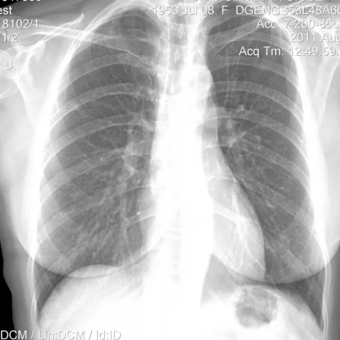
Patient history
History: A 47-year-old female patient presented with fever, chest pain, blood-streaked sputum and right axillary swelling. She had a history of chronic lymphocytic leukaemia and thrombocytopenic purpura, treated with platelet concentrates and high-dose steroids, after which a haematoma developed on the volar surface of her right wrist.
Imaging findings — key points
- Nocardiosis should be considered in immunocompromised patients, including those on high-dose steroids.
- Pulmonary involvement may present with fever, chest pain and blood-streaked sputum.
- Imaging is used to characterise the pulmonary infection and to monitor response during treatment.
- Differential diagnosis includes tuberculosis, aspergillosis, sarcoidosis and neoplastic disease.
- Comparison of pre-treatment and post-treatment radiographs and CT is valuable for assessing resolution.
- Clinical history of chronic lymphocytic leukaemia and steroid therapy raises suspicion for opportunistic infection.
Discussion
This case illustrates the importance of considering nocardiosis in an immunocompromised patient presenting with fever, chest pain and blood-streaked sputum. The history of chronic lymphocytic leukaemia and high-dose steroid therapy predisposes to opportunistic infection. Imaging with radiography and CT supports the diagnosis and enables monitoring during treatment. Differential diagnoses include tuberculosis, aspergillosis, sarcoidosis and neoplastic disease, all of which may produce overlapping pulmonary findings. Microbiological confirmation is essential. Management involves prolonged antimicrobial therapy, and imaging follow-up helps assess response and detect complications.
Differential diagnosis
| Alternative diagnosis | Key features that distinguish it from this case |
|---|---|
| Tuberculosis | May mimic nocardiosis with cavitation and constitutional symptoms; microbiological confirmation is required to distinguish. |
| Aspergillosis | Fungal infection in immunocompromised patients can produce similar pulmonary findings; culture and serology help differentiate. |
| Sarcoidosis | Typically presents with bilateral hilar lymphadenopathy and non-caseating granulomas rather than an acute infective picture. |
| Neoplastic disease | Underlying chronic lymphocytic leukaemia and possible pulmonary involvement must be excluded; imaging and histology aid distinction. |
The confirmed diagnosis itself is not included as an alternative — it is stated separately below.