Advanced TEVAR for Descending Thoracic Aortic Aneurysm
Written and reviewed by RadLearn editorial team · Updated 21 Oct 2026
Figures (8)
Patient history
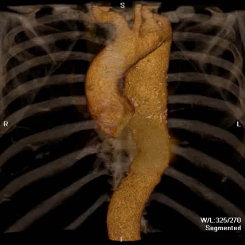
History: A 57-year-old male with recent myocardial infarction and a history of triple aortic bypass surgery in 2008. CT follow-up at 6 months demonstrated enlargement of an aneurysmal dilatation of the descending aorta (diameter: 55 mm) (figures 1 and 2). Because of his relatively young age and the growth pattern of the aneurysm, endovascular treatment was recommended.
Imaging findings — key points
- Descending thoracic aortic aneurysm measuring 55 mm in maximum diameter, with documented enlargement over 6 months.
- CT angiography with 3D volume rendering and MPR MIP reconstructions is used to assess aneurysm morphology and extent.
- Catheter angiography of the aortic arch defines the relationship of the aneurysm to the arch vessels before TEVAR.
- Penetrating aortic ulcer and acute aortic dissection are considered in the differential diagnosis.
- Endovascular treatment (TEVAR) was recommended because of the patient's relatively young age and the growth pattern of the aneurysm.
- Prior triple aortic bypass surgery is relevant background for procedural planning.
Discussion
This case illustrates the role of CT angiography with 3D volume rendering and MPR MIP reconstructions in characterising a descending thoracic aortic aneurysm and planning endovascular repair (TEVAR). The documented enlargement of the aneurysm over 6 months, together with the patient's relatively young age, supported a decision for endovascular treatment. Catheter angiography of the aortic arch remains useful for defining the relationship of the aneurysm to the arch vessels and for stent-graft sizing. Penetrating aortic ulcer and acute aortic dissection are important mimics and were considered in the case analysis. Prior aortic bypass surgery adds complexity to procedural planning. The teaching value lies in the systematic use of cross-sectional and catheter angiography to confirm the diagnosis, exclude mimics and guide a minimally invasive management strategy.
Differential diagnosis
| Alternative diagnosis | Key features that distinguish it from this case |
|---|---|
| Penetrating aortic ulcer | A focal ulcer-like projection within the aortic wall rather than a generalised aneurysmal dilatation; the material lists it as a differential consideration. |
| Acute aortic dissection | Would show an intimal flap and true/false lumen, which are not described in the material; listed as a differential consideration. |
| Mycotic aneurysm | Typically associated with inflammatory changes and clinical infection, not described in the material. |
| Aortic intramural haematoma | Characterised by crescentic mural thickening without a discrete aneurysm, which is not the pattern described. |
The confirmed diagnosis itself is not included as an alternative — it is stated separately below.