Pterygopalatine fossa: a roadmap for disease spread
Written and reviewed by RadLearn editorial team · Updated 20 Oct 2026
Figures (12)
Patient history
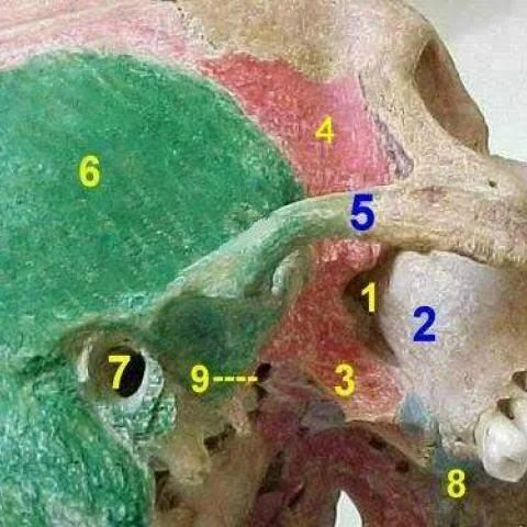
Anatomy of the pterygopalatine fossa.
Imaging findings — key points
- Axial and coronal CT of the skull base shows the pterygopalatine fossa as a small fat-filled space between the posterior wall of the maxillary sinus anteriorly and the pterygoid process of the sphenoid posteriorly.
- The fossa communicates with the middle cranial fossa through the foramen rotundum, which transmits the maxillary division of the trigeminal nerve, and with the nasal cavity through the sphenopalatine foramen.
- Additional communications include the inferior orbital fissure, the pterygoid canal, the palatovaginal canal and the greater palatine canal, all of which are visible on thin-section reformats.
- Sagittal CT and contrast-enhanced axial CT show the internal maxillary artery and its branches within the fossa, together with the pterygopalatine ganglion.
- MRI demonstrates the normal fat signal of the fossa on T1-weighted images and the neural structures as small intermediate-signal structures, with no mass or abnormal enhancement.
Discussion
The pterygopalatine fossa is a small, fat-filled space that serves as a crossroads between the orbit, nasal cavity, oral cavity, middle cranial fossa and infratemporal fossa, and its connections are the key to understanding spread of disease in the head and neck. It contains the maxillary nerve, the pterygopalatine ganglion and branches of the internal maxillary artery, and it communicates through the foramen rotundum, inferior orbital fissure, sphenopalatine foramen and pterygoid canal. On CT and MRI the normal fossa is symmetric and fat-filled, so loss of fat, enlargement of a foramen or abnormal enhancement indicates perineural tumour spread, infection or a vascular lesion. Recognising the normal anatomy on dedicated thin-section imaging is essential before interpreting any asymmetry in this region.
Differential diagnosis
| Alternative diagnosis | Key features that distinguish it from this case |
|---|---|
| Perineural tumour spread | Perineural spread along the maxillary nerve obliterates the normal fat of the pterygopalatine fossa, enlarges the foramen rotundum and shows abnormal enhancement along V2, in contrast with the symmetric fat-filled fossa of normal anatomy. |
| Juvenile nasopharyngeal angiofibroma | Juvenile nasopharyngeal angiofibroma widens the pterygopalatine fossa and erodes the pterygoid plates in an adolescent boy with epistaxis, and it enhances avidly with flow voids. |
| Nerve sheath tumour | A schwannoma or neurofibroma produces a well-defined enhancing mass along the course of V2 with smooth enlargement of the foramen rotundum, rather than normal fat and symmetry. |
| Meningioma of the sphenoid wing | A meningioma extends into the fossa from the middle cranial fossa with dural thickening, hyperostosis and homogeneous enhancement, unlike the normal fat-containing space. |
The confirmed diagnosis itself is not included as an alternative — it is stated separately below.