Juvenile nasopharyngeal angiofibroma
Written and reviewed by RadLearn editorial team · Updated 20 Oct 2026
Figures (9)
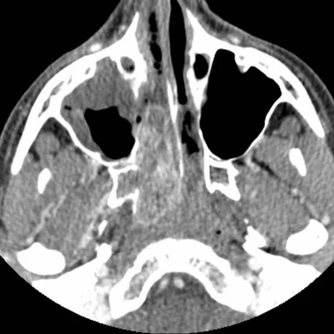
Patient history
A 14-year-old boy with recurrent epistaxis and nasal obstruction.
Imaging findings — key points
- CT with bone window shows a lobulated soft-tissue mass centred on the sphenopalatine foramen and nasopharynx, with widening of the pterygopalatine fossa and erosion of the pterygoid plates.
- There is anterior bowing of the posterior wall of the maxillary sinus and of the medial pterygoid plate, with preservation of the bony margins rather than permeative destruction.
- The mass extends into the nasal cavity and choana, producing nasal obstruction, and it enhances avidly after contrast administration.
- Digital subtraction angiography demonstrates an intense tumour blush supplied predominantly by branches of the internal maxillary artery, with draining veins opacified in the venous phase.
- No cervical lymphadenopathy or intracranial extension beyond the sphenoid sinus is identified.
Discussion
Juvenile nasopharyngeal angiofibroma is a benign but locally aggressive vascular tumour that occurs almost exclusively in adolescent males and typically arises near the sphenopalatine foramen. It presents with recurrent epistaxis and nasal obstruction, and its growth pattern is characterised by expansion along natural pathways with remodelling rather than destruction of bone, producing widening of the pterygopalatine fossa and bowing of the posterior maxillary wall. Contrast-enhanced CT and MRI define the extent, and digital subtraction angiography confirms the diagnosis and allows preoperative embolisation, which reduces operative blood loss. Surgery is the definitive treatment, and recurrence is most likely when the tumour extends into the skull base.
Differential diagnosis
| Alternative diagnosis | Key features that distinguish it from this case |
|---|---|
| Rhabdomyosarcoma | Rhabdomyosarcoma destroys bone in a permeative fashion, grows rapidly and does not produce the smooth widening and bowing of the pterygopalatine fossa seen with an angiofibroma. |
| Nasopharyngeal carcinoma | Nasopharyngeal carcinoma occurs in older adults, causes infiltrative bone destruction and cervical nodal metastases, and is not a highly vascular mass with a blush on angiography. |
| Juvenile nasal polyp | A juvenile polyp is a non-enhancing, fluid-signal mass within the nasal cavity without widening of the pterygopalatine fossa or intense vascularity. |
| Haemangioma | A haemangioma enhances but lacks the characteristic location at the sphenopalatine foramen, the bony remodelling pattern and the arterial supply from the internal maxillary artery. |
The confirmed diagnosis itself is not included as an alternative — it is stated separately below.