Prostate adenocarcinoma, Gleason 7 (3+4)
Written and reviewed by RadLearn editorial team · Updated 22 Oct 2026
Figures (4)
Patient history
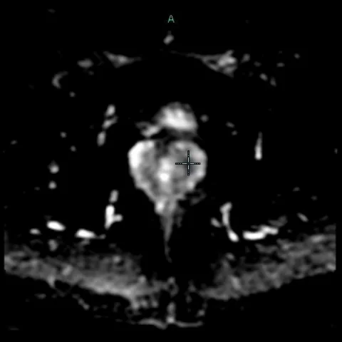
History: Patient with benign prostatic hypertrophy, rising PSA now >12 ng/mL and urinary tract obstruction. Previous colectomy for ulcerative colitis of the colon; the rectum is absent. Imaging work-up included multiparametric MRI and CT.
Imaging findings — key points
- Rising PSA >12 ng/mL with urinary tract obstruction raises suspicion of prostate cancer.
- Diffusion-weighted imaging (ADC map) is key for detecting restricted diffusion in prostate tumours.
- Perfusion imaging (DCE) adds functional information for tumour characterisation.
- Axial T2-weighted imaging provides anatomical detail of the prostate and seminal vesicles.
- Post-contrast T1-weighted SPGR helps assess enhancement patterns.
- CT guidance is used for targeted biopsy; the absent rectum alters the approach.
Discussion
This case illustrates the multiparametric MRI work-up of suspected prostate cancer in a patient with rising PSA and urinary obstruction. The ADC map and perfusion imaging are central to identifying and characterising focal prostatic lesions, while T2-weighted and post-contrast T1-weighted images provide anatomical and enhancement information. A key pitfall is the absent rectum following colectomy for ulcerative colitis of the colon, which alters the usual endorectal or transrectal biopsy approach and may affect image quality. Computer-aided detection (CAD) and CT-guided biopsy were used to target the lesion. The final histology confirmed prostate adenocarcinoma, Gleason 7 (3+4), alongside benign prostatic hypertrophy, prostatic intraepithelial neoplasia and prostatitis. Management implication: accurate Gleason grading guides prognosis and treatment selection.
Differential diagnosis
| Alternative diagnosis | Key features that distinguish it from this case |
|---|---|
| Benign prostatic hypertrophy | Enlarged prostate with transitional zone nodules; usually no marked restricted diffusion or early enhancement. |
| Prostatitis | Diffuse or patchy signal change, often with clinical infection; may show restricted diffusion but usually less focal. |
| Prostatic intraepithelial neoplasia | Histological diagnosis; imaging may be normal or show subtle changes, not a discrete mass. |
| Prostate adenocarcinoma | Focal lesion with restricted diffusion and early enhancement; confirmed by biopsy. |
The confirmed diagnosis itself is not included as an alternative — it is stated separately below.