Primary synovial chondromatosis
Written and reviewed by RadLearn editorial team · Updated 20 Oct 2026
Figures (9)
Patient history
A 40-year-old man presented to his GP in March 2008 with a four-year history of recurrent dislocation of the left shoulder. Physical examination was unremarkable. Outpatient radiographs and MRI were performed, but the patient did not attend follow-up.
Imaging findings — key points
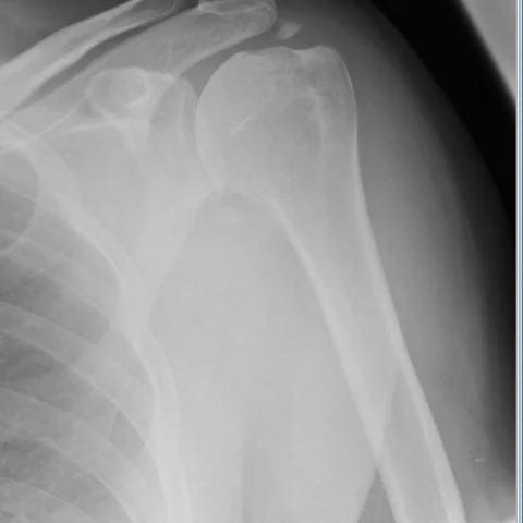
- The AP radiograph of the left shoulder shows multiple well-defined, rounded calcified bodies around the glenohumeral joint, with preservation of the joint space and no erosions.
- Sagittal and coronal proton density fat-saturated MRI show numerous intra-articular and periarticular nodules that are isointense to cartilage and hyperintense on T2-weighted images, with a ring-and-arc pattern of calcification.
- The nodules are distributed within the joint recesses and subacromial bursa, and a moderate joint effusion is present.
- There is no bone marrow invasion, soft-tissue mass or rotator cuff tear, and the glenoid and humeral head show no destructive change.
- Follow-up radiographs show an increase in the number and size of the calcified bodies.
Discussion
Primary synovial chondromatosis is a benign metaplastic disorder in which synovial cells form multiple cartilaginous nodules that may calcify and ossify within the joint, most often affecting the knee, hip and shoulder. It presents with pain, swelling and mechanical symptoms such as locking or recurrent dislocation, and imaging shows multiple intra-articular bodies with a ring-and-arc calcification pattern and preserved joint space. The main differentials are degenerative joint disease, calcific tendinitis and rheumatoid arthritis, none of which produce multiple chondral loose bodies. Treatment is arthroscopic or open synovectomy with removal of the loose bodies, and recurrence is possible, especially after incomplete synovectomy.
Differential diagnosis
| Alternative diagnosis | Key features that distinguish it from this case |
|---|---|
| Degenerative joint disease | Osteoarthrosis shows joint space narrowing, subchondral sclerosis and marginal osteophytes, and the calcified bodies are not multiple loose bodies within the joint recesses. |
| Calcific tendinitis | Calcific tendinitis produces amorphous calcification within the rotator cuff tendon, not multiple well-defined intra-articular chondral bodies. |
| Rheumatoid arthritis | Rheumatoid arthritis causes marginal erosions, uniform joint space loss and pannus enhancement, without multiple calcified loose bodies. |
| Osteochondritis dissecans | Osteochondritis dissecans shows a donor site in the articular surface with a detached osteochondral fragment, rather than diffuse synovial chondromatosis. |
The confirmed diagnosis itself is not included as an alternative — it is stated separately below.