Adenomyoepithelioma
Written and reviewed by RadLearn editorial team · Updated 20 Oct 2026
Figures (19)
Patient history
Patient with a history of left breast malignancy 23 years ago, treated with mastectomy, left axillary lymph node dissection, and radiotherapy. Recently diagnosed with a second malignant nodule in the left breast, metastatic right axillary lymphadenopathy, and a pseudonodular area in the right breast that has been known and stable for years.
Imaging findings — key points
- Mammography of the right breast shows a round-to-oval, well-circumscribed mass of intermediate density without spiculation, kinking of the surrounding tissue or associated microcalcifications.
- Serial examinations over several years show that this mass has remained stable in size and morphology, a feature that argues strongly against a malignant lesion.
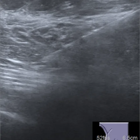
- Ultrasound demonstrates a solid, circumscribed, homogeneous hypoechoic nodule with lobulated margins and no posterior acoustic shadowing.
- MRI shows a well-defined mass with smooth margins, isointense signal on T1-weighted images, mildly heterogeneous hyperintensity on T2-weighted images and avid heterogeneous enhancement without washout kinetics.
- The separate left breast lesion and the right axillary nodes show malignant features with irregular margins and washout kinetics, and are clearly distinct from the stable right-breast nodule.
Discussion
Adenomyoepithelioma of the breast is an uncommon tumour that contains both epithelial and myoepithelial cells and usually presents as a well-circumscribed, mobile mass. It has no specific imaging signature because mammography and ultrasound show a circumscribed solid lesion and MRI shows avid enhancement that can mimic malignancy, so imaging follow-up or biopsy is usually required. Malignant transformation is rare, but complete excision is advised because of a tendency to local recurrence when the lesion is incompletely removed.
Differential diagnosis
| Alternative diagnosis | Key features that distinguish it from this case |
|---|---|
| Breast adenosis | Presents with clustered punctate calcifications or a diffuse heterogeneous pattern without a discrete stable circumscribed mass, and lacks the biphasic epithelial and myoepithelial proliferation of adenomyoepithelioma. |
| Hamartoma or fibroadenolipoma | Contains a mixture of fat, fibrous and glandular tissue within a pseudocapsule and shows fat-density components on mammography, which are absent here. |
| Ductal carcinoma in situ | Typically manifests as segmental linear-branching microcalcifications or clumped non-mass enhancement on MRI, rather than a long-standing stable circumscribed solid nodule. |
The confirmed diagnosis itself is not included as an alternative — it is stated separately below.