Microcystic calcifications of fat necrosis
Written and reviewed by RadLearn editorial team · Updated 20 Oct 2026
Figures (20)
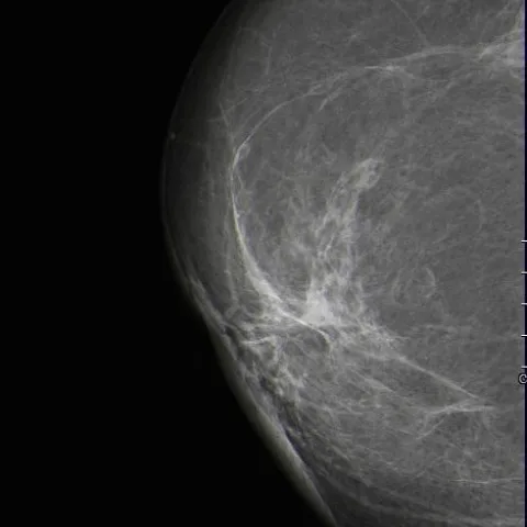
Patient history
A 52-year-old female patient presented with a painful mass in the left breast. She had a history of bilateral breast reduction surgery one year prior (2012). Clinical examination revealed a hard mass in the upper outer quadrant of the left breast.
Imaging findings — key points
- Mammography shows an ill-defined area of mixed density in the upper outer quadrant of the left breast containing round lucent-centred oil cysts, in a patient with previous reduction mammoplasty.
- Within this area there are clusters of microcystic calcifications with a partially lucent centre, the characteristic appearance of fat necrosis.
- Sequential mammograms obtained in 2013, 2015 and 2016 document evolution of the density and progressive development of the calcifications, without a spiculated mass or linear-branching calcification.
- Ultrasound shows an ill-defined mixed-echogenicity lesion with internal echogenic fat and focal shadowing from calcification, without a complex cyst containing septa or a vascular mural nodule.
- MRI shows a heterogeneous area containing fat-signal components (oil cysts) that are hyperintense on T1- and T2-weighted images, with rim enhancement and no washout.
Discussion
Fat necrosis of the breast is a benign condition that commonly follows surgery, trauma or radiotherapy and may develop after reduction mammoplasty. It produces a spectrum of imaging appearances including oil cysts, coarse rim calcifications and, as in this case, clusters of microcystic calcifications, all reflecting the underlying fat and its subsequent fibrosis. The presence of a lucent centre within the calcifications and of fat signal on MRI allows fat necrosis to be distinguished from the suspicious calcifications of ductal carcinoma in situ, although biopsy may still be needed when the pattern is equivocal.
Differential diagnosis
| Alternative diagnosis | Key features that distinguish it from this case |
|---|---|
| Seroma | Appears as a well-defined unilocular anechoic collection without internal echoes, septa or fat signal, and does not contain calcification on mammography. |
| Haematoma | Evolves over time with changing internal echoes and a haemosiderin rim, and in the subacute phase is hyperintense on T1-weighted MRI without a fat-fluid or oil cyst appearance. |
| Complex cyst with septations | Shows enhancing internal septa and thickened walls with internal vascularity, and lacks the fat-density lucency and lucent-centred calcifications of fat necrosis. |
| Intracystic papillary carcinoma | Presents as a complex cystic lesion with a solid mural nodule that shows internal vascularity, in contrast to the avascular wall calcifications and oil cysts of fat necrosis. |
The confirmed diagnosis itself is not included as an alternative — it is stated separately below.