Pedunculated Intracavitary Left Ventricular Lipoma
Written and reviewed by RadLearn editorial team · Updated 22 Oct 2026
Figures (4)
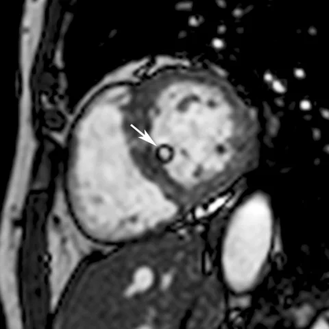
Patient history
History: A 65-year-old asymptomatic woman with a history of hypertension was found to have a left ventricular mass on routine transthoracic echocardiography. Cardiac MRI was performed to further characterise the mass.
Imaging findings — key points
- Cardiac lipomas are rare primary cardiac tumours; approximately 75% of primary cardiac tumours are benign and 25% malignant.
- Most benign cardiac tumours are myxomas; most of the remainder are lipomas, papillary fibroelastomas and rhabdomyomas.
- Cardiac lipomas account for 8% of all cardiac tumours and occur at any age and in both sexes.
- They are well-encapsulated tumours composed of mature adipocytes, distinguished from unencapsulated hypertrophic lipomatosis of the atrial septum.
- 50% arise intracavitarily and subendocardially, 25% intramyocardially, and 25% epicardially and extracavitarily; the right atrium is the commonest site.
- Echocardiography is non-specific, showing a uniform, hyperechoic mass; cardiac MRI is necessary to characterise location, extent, tissue composition, attachment and functional impact.
- Lipomas show uniform hyperintensity on T1-weighted imaging, slightly lower hyperintensity on T2-weighted imaging, and signal loss on fat-saturation sequences; they do not enhance with contrast.
- SSFP cine MRI is particularly useful for assessing mobile tumours such as pedunculated lipomas.
Discussion
Primary cardiac tumours are rare, and approximately 75% are benign; after myxoma, lipoma is among the commonest benign types, accounting for 8% of all cardiac tumours. Cardiac lipomas are well-encapsulated tumours of mature adipocytes and must be distinguished from unencapsulated hypertrophic lipomatosis of the atrial septum. Half arise intracavitarily and subendocardially, a quarter intramyocardially and a quarter epicardially or extracavitarily; the right atrium is the commonest site, but they also occur in the pericardium, left atrium, valves, pulmonary veins, coronary arteries and ventricles. Most patients, as in this case, are asymptomatic and the tumour is incidental. Echocardiography shows a non-specific uniform hyperechoic mass, so cardiac MRI is required to characterise location, extent, tissue composition, attachment and functional impact. Lipomas are uniformly hyperintense on T1-weighted imaging, slightly hyperintense on T2-weighted imaging and lose signal on fat saturation, and they do not enhance. SSFP cine imaging is valuable for mobile pedunculated tumours. Symptomatic lesions are treated by surgical excision.
Differential diagnosis
| Alternative diagnosis | Key features that distinguish it from this case |
|---|---|
| Cardiac myxoma | Myxomas are the commonest benign cardiac tumour but do not show fat signal loss on fat-saturation sequences and typically enhance with contrast. |
| Fibroblastoma | Fibroblastomas are fibrous rather than fatty tumours and would not demonstrate uniform fat signal with signal loss on fat saturation. |
| Hypertrophic lipomatosis of the atrial septum | This is unencapsulated deposition of mature and fetal adipose tissue, most commonly in the atrial septum, unlike the well-encapsulated intracavitary lipoma seen here. |
The confirmed diagnosis itself is not included as an alternative — it is stated separately below.