Extramedullary haematopoiesis
Written and reviewed by RadLearn editorial team · Updated 20 Oct 2026
Figures (8)
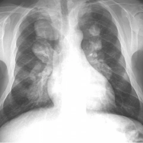
Patient history
A 31-year-old man with known beta-thalassaemia and a history of diffuse chest pain, dyspnoea and migraine was referred to our department for a standard chest assessment, followed by CT of the chest and brain. One year later, MRI of the cervicothoracic spine was performed for paraesthesia at the T3-T4 level and cord compression.
Imaging findings — key points
- The PA chest radiograph shows bilateral, well-defined paravertebral soft-tissue masses in the lower thorax, with widening of several ribs and a diffuse increase in bone density.
- CT of the chest demonstrates multiple broad-based, lobulated paravertebral masses of homogeneous soft-tissue attenuation, without bone destruction or calcification, and the adjacent ribs are expanded with coarse trabeculation.
- Head CT with intravenous contrast shows widening of the diploic space with a hair-on-end appearance of the calvarium, and the bone window confirms diffuse marrow expansion.
- Sagittal MRI of the cervicothoracic spine shows a large epidural and paravertebral mass that is isointense on T1-weighted images and hyperintense on T2-weighted images, compressing the cord at the T3-T4 level.
- The masses do not invade the bone and show only mild, heterogeneous enhancement, consistent with haematopoietic tissue rather than a neoplasm.
Discussion
Extramedullary haematopoiesis is the formation of blood cells outside the marrow cavity when chronic anaemia, most often thalassaemia, sickle cell disease or myelofibrosis, overwhelms marrow capacity. It typically produces bilateral paravertebral masses in the lower thorax, rib expansion, a hair-on-end skull and, when epidural, spinal cord compression, and the masses consist of hyperplastic haematopoietic tissue rather than tumour. The imaging diagnosis rests on the combination of chronic anaemia, bone marrow expansion and non-destructive paravertebral masses, and biopsy should be avoided because of the risk of bleeding. Treatment is directed at the underlying anaemia, with radiotherapy or surgery reserved for symptomatic cord compression.
Differential diagnosis
| Alternative diagnosis | Key features that distinguish it from this case |
|---|---|
| Neurogenic tumour | A schwannoma or neurofibroma is a well-defined paravertebral mass that often extends through a neural foramen and enhances intensely, without diffuse marrow expansion or rib widening. |
| Lymphoma | Lymphoma produces bulky, homogeneous lymph node masses that may encase vessels and show restricted diffusion, and it is not associated with generalised marrow hyperplasia. |
| Metastasis | Metastatic disease destroys bone, produces lytic or sclerotic vertebral lesions and is usually painful and progressive, whereas these masses preserve the bone and expand the marrow. |
| Paravertebral abscess | An abscess shows rim enhancement with central fluid, often with discitis and endplate destruction, and the patient has fever and raised inflammatory markers. |
The confirmed diagnosis itself is not included as an alternative — it is stated separately below.