Parenchymal Neurocysticercosis
Written and reviewed by RadLearn editorial team · Updated 21 Oct 2026
Figures (5)
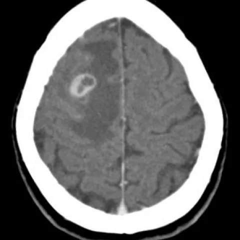
Patient history
History: A 35-year-old woman who recently immigrated from Russia to Belgium presented with two episodes of left-sided focal motor seizures beginning in the upper limb and spreading to the face and lower limb. She reported mild headache over the past week. Neurological examination showed weakness of the left limbs.
Imaging findings — key points
- Focal parenchymal lesions with ring or nodular enhancement on post-contrast CT and gadolinium-enhanced T1WI.
- Surrounding oedema is best appreciated as hyperintensity on FLAIR and low attenuation on CT.
- DWI/ADC show no restricted diffusion, helping to exclude a pyogenic brain abscess.
- Lesion distribution and appearance favour the colloidal-vesicular or granulomatous stage of neurocysticercosis.
- Tuberculoma and metastatic nodule remain important mimics and require clinical and laboratory correlation.
- Contrast-enhanced MRI is the examination of choice for characterising the lesions and assessing oedema.
Discussion
Parenchymal neurocysticercosis is the most common parasitic infection of the central nervous system and a frequent cause of focal seizures in patients from endemic regions. The diagnosis rests on recognising the stage-specific imaging appearances: vesicular lesions with a visible scolex, colloidal-vesicular lesions with ring enhancement and oedema, and granulomatous or calcified nodules. The key pitfall is mistaking a ring-enhancing lesion for a pyogenic brain abscess; DWI/ADC is invaluable because abscess cavities restrict diffusion whereas cysticercal cysts generally do not. Tuberculoma and metastatic nodule are the other main mimics and require correlation with history, laboratory tests and follow-up imaging. Contrast-enhanced MRI is the examination of choice for characterising the lesions and assessing oedema. Management implication: accurate staging on imaging guides antiparasitic and corticosteroid therapy and helps avoid unnecessary surgery.
Differential diagnosis
| Alternative diagnosis | Key features that distinguish it from this case |
|---|---|
| Brain abscess | A pyogenic brain abscess typically shows marked restricted diffusion (low ADC) within the cavity, which is not demonstrated here. |
| Tuberculoma | Tuberculomas may show ring enhancement and oedema but often have a target appearance and may show restricted diffusion; clinical and epidemiological context helps differentiate. |
| Metastatic nodule | Metastases are usually multiple, at the grey-white junction, with more prominent vasogenic oedema and often a known primary tumour. |
| Primary malignant brain tumour | Primary tumours usually show mass effect, irregular enhancement and restricted diffusion; the small focal lesions here are less suggestive. |
| Other parasitic infection (e.g. toxoplasmosis) | Toxoplasmosis typically occurs in immunocompromised patients with multiple ring-enhancing lesions and restricted diffusion; the clinical context differs. |
The confirmed diagnosis itself is not included as an alternative — it is stated separately below.