Olfactory groove meningioma
Written and reviewed by RadLearn editorial team · Updated 20 Oct 2026
Figures (17)
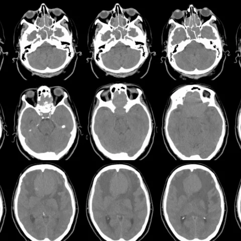
Patient history
Subacute clinical picture of headache, gait disturbance (falls at home) and apathy with confusion, worsening in the week before neuroimaging and diagnosis. Long-standing anosmia was noted retrospectively. There were no complaints of nasal obstruction, rhinorrhoea or epistaxis. Personal history of depression.
Imaging findings — key points
- Unenhanced CT shows a large, well-defined, iso- to slightly hyperdense extra-axial mass in the olfactory groove, with a broad dural base along the planum sphenoidale and adjacent hyperostosis.
- Contrast-enhanced CT and MRI demonstrate intense, homogeneous enhancement of the mass, with a dural tail extending posteriorly along the falx and no central necrosis.
- The mass is iso- to slightly hypointense on T1-weighted images, iso- to hyperintense on T2-weighted images and shows a cerebrospinal fluid cleft at its margin, confirming its extra-axial location.
- There is upward displacement of the frontal lobes with surrounding vasogenic oedema and mild subfalcine herniation, and the olfactory tracts are not separately identifiable.
- MR spectroscopy shows an elevated choline peak, reduced N-acetylaspartate and a prominent lipid or lactate peak, supporting a meningothelial tumour rather than a intra-axial glioma.
Discussion
Olfactory groove meningioma is a benign extra-axial tumour arising from arachnoid cap cells along the cribriform plate and planum sphenoidale, and it typically grows slowly, reaching a large size before producing symptoms. Progressive anosmia is often long standing and overlooked, while frontal lobe compression causes apathy, confusion, gait disturbance and headache. Imaging is characteristic when a broad-based, homogeneously enhancing extra-axial mass with a dural tail and hyperostosis is seen; the main differential is a nasal or skull-base malignancy, which tends to erode bone rather than thicken it. Surgery is curative in most cases, and the extent of resection depends on involvement of the anterior cerebral arteries and optic apparatus.
Differential diagnosis
| Alternative diagnosis | Key features that distinguish it from this case |
|---|---|
| Olfactory neuroblastoma | An olfactory neuroblastoma arises in the nasal cavity, destroys the cribriform plate and contains calcification, whereas an olfactory groove meningioma is centred intracranially with hyperostosis and homogeneous enhancement. |
| Skull base metastasis | A metastasis usually causes bone destruction, heterogeneous enhancement and marked oedema, and is often multiple or associated with a known primary tumour. |
| Sinonasal carcinoma | Sinonasal carcinoma presents with nasal obstruction, bone erosion and mucosal disease, and does not show the broad dural base and dural tail of a meningioma. |
| Dural metastasis or granuloma | Dural deposits are often multifocal and nodular with a known systemic disease, and lack the uniform enhancement and hyperostosis of a meningioma. |
The confirmed diagnosis itself is not included as an alternative — it is stated separately below.