Male breast cancer, synchronous tumours, bilateral
Written and reviewed by RadLearn editorial team · Updated 20 Oct 2026
Figures (17)
Patient history
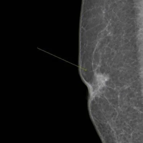
A 59-year-old male presented to our hospital with a palpable mass in the right breast. Physical examination revealed a fixed, painless mass in the right breast and a similar mass in the contralateral breast. There was no family history of breast cancer, solid organ tumours, or hormone therapy.
Imaging findings — key points
- Bilateral mammography (craniocaudal and mediolateral oblique views) demonstrates a high-density, irregular, spiculated mass in the right breast and a similar second mass in the left breast.
- Both masses are eccentric to the nipple-areolar complex rather than centred posterior to it, and neither shows the flame-shaped glandular tails typical of gynaecomastia.
- Ultrasound demonstrates irregular hypoechoic solid masses with non-parallel orientation in both breasts, with abnormal axillary lymph nodes showing cortical thickening and loss of the fatty hilum.
- Histopathology and immunohistochemistry of both lesions confirm invasive carcinoma, and sentinel lymph node biopsy of the right axilla provides nodal staging.
- The skin and subcutaneous fat are otherwise unremarkable, and there is no associated abscess or fluid collection.
Discussion
Male breast cancer is uncommon, and the presence of synchronous bilateral tumours in one patient is rare. Because the male breast contains only ducts and fat, a solid eccentric spiculated retroareolar mass, particularly when it is separate from the nipple and accompanied by axillary adenopathy, is suspicious for carcinoma and must be distinguished from gynaecomastia. Mammography and ultrasound of both breasts are therefore required for staging, and histopathology with immunohistochemistry confirms the diagnosis and guides treatment.
Differential diagnosis
| Alternative diagnosis | Key features that distinguish it from this case |
|---|---|
| Gynaecomastia | Produces flame-shaped, centrally located retroareolar glandular tissue symmetrical about the nipple, without a spiculated eccentric mass, microcalcifications or axillary adenopathy. |
| Abscess | Presents as a tender fluctuant complex fluid collection with surrounding inflamed fat and systemic symptoms rather than a solid spiculated mass. |
| Breast sarcoma | Is a large heterogeneous mesenchymal mass without an eccentric subareolar location or the epithelial immunohistochemical profile of primary breast carcinoma. |
| Metastatic disease to the breast | Usually arises from the contralateral breast, melanoma or lymphoma, presents as one or more circumscribed masses, and requires identification of a primary tumour elsewhere. |
The confirmed diagnosis itself is not included as an alternative — it is stated separately below.