Infiltrating ductal carcinoma of the right breast
Written and reviewed by RadLearn editorial team · Updated 20 Oct 2026
Figures (8)
Patient history
A 53-year-old male patient was referred to our hospital's breast clinic with a palpable mass in the right breast. Physical examination revealed a painless, fixed, unilateral mass in the upper outer quadrant of the right breast. No history of malignancy.
Imaging findings — key points
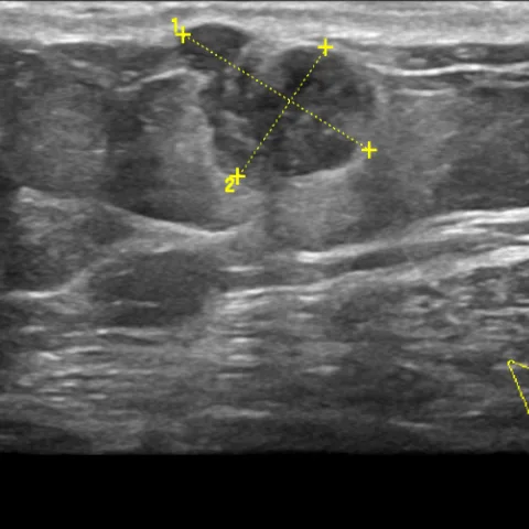
- B-mode ultrasound depicts a suspicious hypoechoic solid mass in the right upper outer quadrant with irregular, indistinct margins, non-parallel orientation and posterior acoustic shadowing.
- Colour Doppler and B-flow imaging show internal vessels within the mass, indicating hypervascularity.
- Strain elastography and shear-wave elastography demonstrate markedly increased stiffness of the lesion, supporting malignancy.
- Digital mammography (RMLO and RCC views) shows a dense, spiculated mass in the upper outer quadrant of the right breast without coarse or pleomorphic calcification.
- The mass is eccentric to the nipple rather than subareolar, a distribution that favours male breast carcinoma over gynaecomastia.
- Ultrasound-guided hook-wire localisation was performed to guide surgical excision.
Discussion
Male breast carcinoma is uncommon, accounting for less than 1% of all breast cancers, and invasive ductal carcinoma is by far its most frequent histological type. A painless, fixed, eccentric unilateral mass in a 53-year-old man is highly suspicious, so the same triple assessment used in women (clinical examination, mammography with ultrasound, and tissue sampling) should be applied. Masses that are hypoechoic and irregular with internal vascularity and markedly increased stiffness on elastography should be regarded as malignant until proven otherwise, and because of the small volume of the male breast, mastectomy with sentinel node assessment is generally required for local control.
Differential diagnosis
| Alternative diagnosis | Key features that distinguish it from this case |
|---|---|
| Gynaecomastia (nodular type) | Presents as a subareolar, fan-shaped or flame-shaped retroareolar density that is symmetric and shows neither spiculation nor internal vascularity, unlike the eccentric spiculated mass seen here. |
| Metastasis to the breast | Usually appears as multiple superficial, round, circumscribed masses without spiculation and without an in-situ component, typically in a patient with a known extramammary malignancy. |
| Myofibroblastoma | A benign spindle-cell tumour that is well-circumscribed and oval with smooth margins, showing no spiculation, no posterior shadowing and no internal vascularity. |
| Liposarcoma and other soft-tissue sarcomas | Characteristically contain macroscopic fat or lipomatous components visible on mammography and MRI, which are absent in this lesion. |
| Diabetic mastopathy | Occurs in insulin-dependent diabetic men as ill-defined, very hypoechoic masses with dense fibrous stroma that show little or no enhancement on MRI. |
The confirmed diagnosis itself is not included as an alternative — it is stated separately below.