Mounier-Kuhn syndrome (tracheobronchomegaly)
Written and reviewed by RadLearn editorial team · Updated 20 Oct 2026
Figures (7)
Patient history
A 42-year-old man attended the outpatient respiratory clinic with a long-standing history of productive cough and recurrent chest infections.
Imaging findings — key points
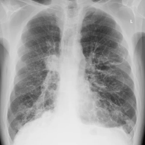
- The chest radiograph shows a widened, air-filled trachea and increased bronchovascular markings in both lower zones, with no focal consolidation.
- CT of the chest in the lung window demonstrates a markedly dilated trachea with a transverse diameter greater than 30 mm at the level of the aortic arch.
- The main bronchi are also dilated, exceeding 20 mm on the right and 18 mm on the left, and there are multiple small diverticula arising from the tracheal and bronchial walls.
- There is bilateral lower-lobe bronchiectasis with bronchial wall thickening, mucoid impaction and recurrent infection-related scarring.
- Coronal reconstruction confirms diffuse tracheobronchomegaly with a smooth, thinned wall and no focal mass or stricture.
Discussion
Mounier-Kuhn syndrome, or tracheobronchomegaly, is a rare disorder characterised by marked dilatation of the trachea and main bronchi due to atrophy of the elastic tissue and smooth muscle, often with an inherited component. Patients present with chronic productive cough, recurrent chest infections and bronchiectasis, and CT is diagnostic when the trachea exceeds 30 mm, the right main bronchus 20 mm and the left 18 mm, with diverticula and bronchial dilatation. The main differentials are other causes of bronchiectasis and Williams-Campbell syndrome, which lacks the diffuse tracheal enlargement. There is no specific treatment; management focuses on airway clearance, physiotherapy and prompt treatment of infection.
Differential diagnosis
| Alternative diagnosis | Key features that distinguish it from this case |
|---|---|
| Bronchiectasis | Bronchiectasis alone dilates the peripheral bronchi but does not produce diffuse tracheal enlargement beyond 30 mm or tracheal diverticula. |
| Williams-Campbell syndrome | Williams-Campbell syndrome is a congenital deficiency of bronchial cartilage that causes cystic bronchiectasis, whereas Mounier-Kuhn syndrome is defined by tracheobronchomegaly with diverticula. |
| Tracheomalacia | Tracheomalacia causes excessive expiratory collapse of a normal-calibre trachea and is best shown on dynamic imaging, without diffuse enlargement. |
| Allergic bronchopulmonary mycosis | ABPA causes central bronchiectasis with mucoid impaction, high IgE and eosinophilia, but the trachea remains of normal calibre. |
The confirmed diagnosis itself is not included as an alternative — it is stated separately below.