Intracranial tuberculous granuloma (tuberculoma)
Written and reviewed by RadLearn editorial team · Updated 20 Oct 2026
Figures (19)
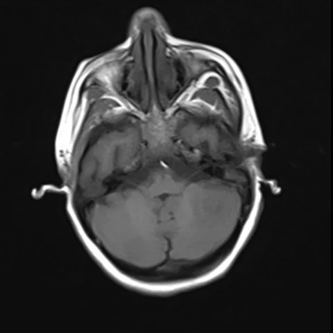
Patient history
A 19-year-old woman presented with one month of headache, vomiting and low-grade fever. She had a history of pulmonary tuberculosis two years earlier and had defaulted on treatment.
Imaging findings — key points
- T1-weighted images show an isointense to hypointense parenchymal lesion, and T2-weighted images show a predominantly iso- to hypointense centre with a hyperintense rim, reflecting caseous necrosis with a cellular granulomatous capsule.
- FLAIR images demonstrate surrounding vasogenic oedema that is disproportionately mild for the size of the lesion, together with hyperintense sulcal and basal meninges.
- DWI shows no restricted diffusion of the central core, an important feature that distinguishes a tuberculoma from a pyogenic abscess.
- Post-contrast T1-weighted images show ring or nodular enhancement, with a target-like appearance produced by a central non-enhancing area and an enhancing rim, and additional smaller enhancing nodules may be present.
- Mild mass effect and mild ventriculomegaly are present, without midline shift, and no calcification is seen on the available sequences.
Discussion
Intracranial tuberculoma is a granulomatous mass that results from haematogenous spread of Mycobacterium tuberculosis and is common in endemic regions, particularly in patients with a history of pulmonary tuberculosis or poor treatment adherence. The lesion evolves from a non-caseating granuloma to a caseating mass with a collagenous capsule, which explains the T2 hypointensity of the core, the target-like enhancement and the absence of central diffusion restriction. Recognising these features allows a tuberculoma to be distinguished from pyogenic abscess and from neurocysticercosis, both of which have different treatment. The diagnosis is supported by a compatible history, a positive tuberculin test or interferon-gamma release assay, and response to antituberculous therapy; biopsy is reserved for atypical lesions.
Differential diagnosis
| Alternative diagnosis | Key features that distinguish it from this case |
|---|---|
| Neurocysticercosis | A cysticercal cyst is usually smaller, contains a mural scolex, shows no oedema when calcified and does not have the caseous, non-restricted core of a tuberculoma. |
| Pyogenic brain abscess | A pyogenic abscess shows marked restricted diffusion of its central pus with a thin, smooth enhancing capsule, whereas a tuberculoma has no central diffusion restriction and a thicker irregular rim. |
| Cerebral toxoplasmosis | Toxoplasmosis occurs in immunocompromised patients and typically produces multiple basal ganglia lesions with homogeneous or ring enhancement and the asymmetrical target sign. |
| Central nervous system lymphoma | Lymphoma is usually periventricular, homogeneous, markedly diffusion-restricted and shows intense uniform enhancement without central necrosis in immunocompetent patients. |
The confirmed diagnosis itself is not included as an alternative — it is stated separately below.