Bochdalek hernia
Written and reviewed by RadLearn editorial team · Updated 20 Oct 2026
Figures (7)
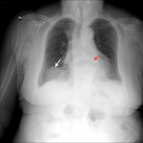
Patient history
An elderly lady, widowed for several years and with no children or relatives, was admitted to a retirement home with the first symptoms of dementia. A chest radiograph performed during a routine check showed an abnormality.
Imaging findings — key points
- The chest radiograph shows a large, smooth, rounded opacity in the left lower hemithorax, obscuring the medial aspect of the left hemidiaphragm and containing subtle gas lucencies.
- Multidetector CT in coronal and sagittal planes demonstrates a defect in the posterior aspect of the left hemidiaphragm with herniation of retroperitoneal fat and bowel into the pleural cavity.
- The herniated contents pass through the Bochdalek foramen and show a collar sign at the diaphragmatic defect, with mass effect on the adjacent lung.
- The left kidney remains in a normal position, and there is no pleural effusion or lung mass.
- The lung parenchyma adjacent to the hernia shows compressive atelectasis but no consolidation or nodule.
Discussion
Bochdalek hernia is a congenital posterolateral diaphragmatic defect that usually presents in neonates with respiratory distress, but a minority remain asymptomatic until adulthood, when they are found incidentally or present with abdominal pain, obstruction or strangulation. CT is definitive, showing a defect in the posterior diaphragm with herniation of fat, omentum, bowel, spleen or kidney into the thorax and a collar sign at the neck of the hernia. The main differentials are a lung or pleural mass, a diaphragmatic lipoma and eventration, all of which lack a true defect. Symptomatic or complicated hernias require surgical repair, and even asymptomatic adult cases are often repaired because of the risk of strangulation.
Differential diagnosis
| Alternative diagnosis | Key features that distinguish it from this case |
|---|---|
| Pulmonary or pleural tumour | A lung or pleural tumour is a solid mass that enhances and may invade the chest wall, without a diaphragmatic defect or herniated fat and bowel. |
| Diaphragmatic lipoma | A diaphragmatic lipoma is a fat-containing mass confined to the diaphragm without a defect or herniation of abdominal contents. |
| Diaphragmatic eventration | Eventration is a smooth elevation of an intact hemidiaphragm without a focal defect or herniated viscera, and the diaphragm remains continuous. |
| Pleural effusion or empyema | A loculated effusion is fluid density with pleural enhancement and no connection to the abdominal cavity. |
The confirmed diagnosis itself is not included as an alternative — it is stated separately below.