Malignant phyllodes tumour of the breast
Written and reviewed by RadLearn editorial team · Updated 20 Oct 2026
Figures (11)
Patient history
A 49-year-old patient presented to our hospital with a left breast mass that had been growing over the past 4 years. Physical examination revealed a hard mass causing skin ulceration. No axillary lymphadenopathy was detected clinically.
Imaging findings — key points
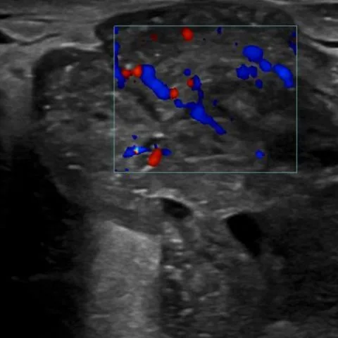
- Ultrasound shows a large heterogeneous mass with both solid and cystic components occupying almost the entire breast, with marked internal vascularity on Doppler.
- The mass extends to the pectoralis muscle and cannot be separated from it on clinical examination.
- CT shows a multilobulated mass with necrotic and heterogeneous density areas occupying the whole left breast.
- The mass infiltrates the pectoralis muscle without invading other chest wall structures.
- No distant metastatic disease is identified on CT.
- The resected specimen measured 24 x 18 x 16 cm on final histology.
Discussion
Phyllodes tumours are fibroepithelial neoplasms arising from both epithelial and stromal breast tissue and are classified as benign, borderline or malignant according to histological features, with the epithelial elements distinguishing them from pure stromal sarcomas. Clinically they present as large, smooth, painless masses showing an initial slow growth phase followed by rapid acceleration; features such as skin ulceration, chest wall invasion or metastasis are uncommon but indicate aggressive behaviour. Imaging shows a circumscribed lobulated mass with solid and cystic components, necrosis and prominent vascularity, and MRI may show cystic areas and heterogeneous delayed enhancement. Core biopsy demonstrating stromal overgrowth allows grading and guides treatment, which is wide local excision with margins greater than 1 cm, or mastectomy for large or malignant tumours.
Differential diagnosis
| Alternative diagnosis | Key features that distinguish it from this case |
|---|---|
| Fibroadenoma | Fibroadenoma is a smaller (usually less than 3 cm), well-circumscribed, homogeneous hypoechoic oval mass with a thin capsule, without the rapid growth, necrosis, cystic change or chest wall infiltration seen here. |
| Primary breast sarcoma | Primary sarcoma also forms a large heterogeneous mass but lacks the epithelial component and leaf-like architecture of a phyllodes tumour, which is confirmed on histology and immunohistochemistry. |
| Locally advanced breast carcinoma | Invasive carcinoma usually shows spiculated margins with suspicious microcalcifications and skin or nipple changes and does not typically produce the hugely lobulated, cystic-necrotic mass with rapid interval growth seen in a malignant phyllodes tumour. |
The confirmed diagnosis itself is not included as an alternative — it is stated separately below.